. 2009 Oct 1;334(1):213-23.
doi: 10.1016/j.ydbio.2009.07.017. Epub 2009 Jul 22.

Loss of Dnmt1 catalytic activity reveals multiple roles for DNA methylation during pancreas development and regeneration

Affiliations

Loss of Dnmt1 catalytic activity reveals multiple roles for DNA methylation during pancreas development and regeneration

Ryan M Anderson et al. Dev Biol. .

Abstract

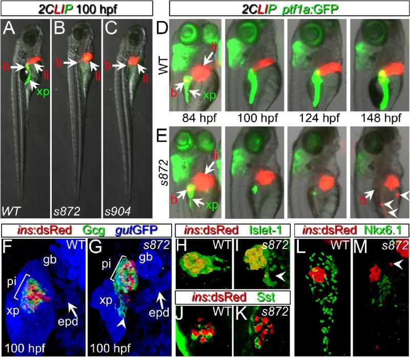
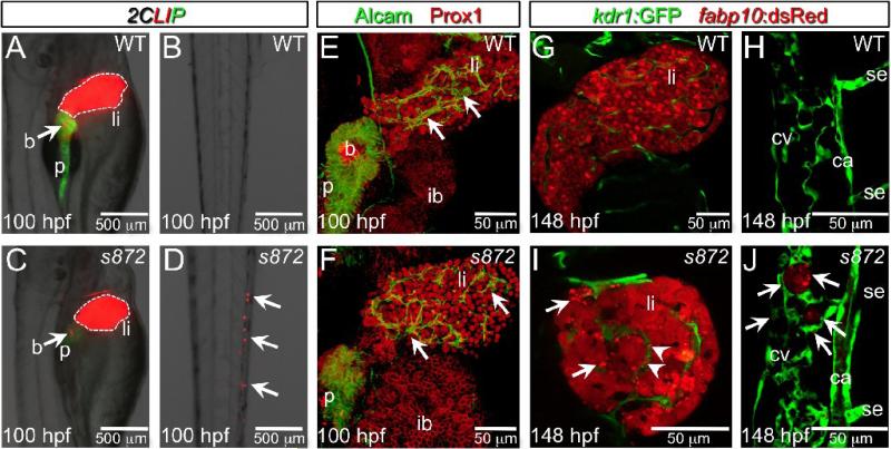
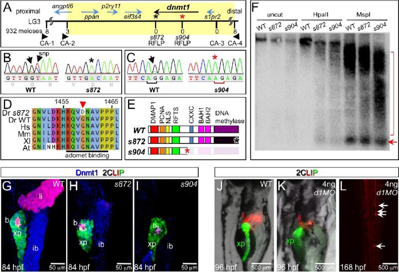
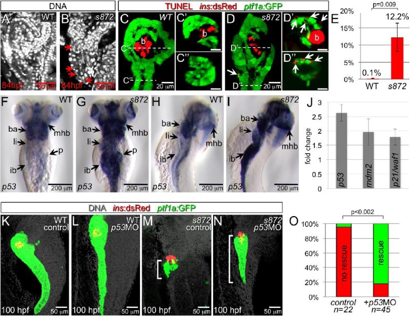
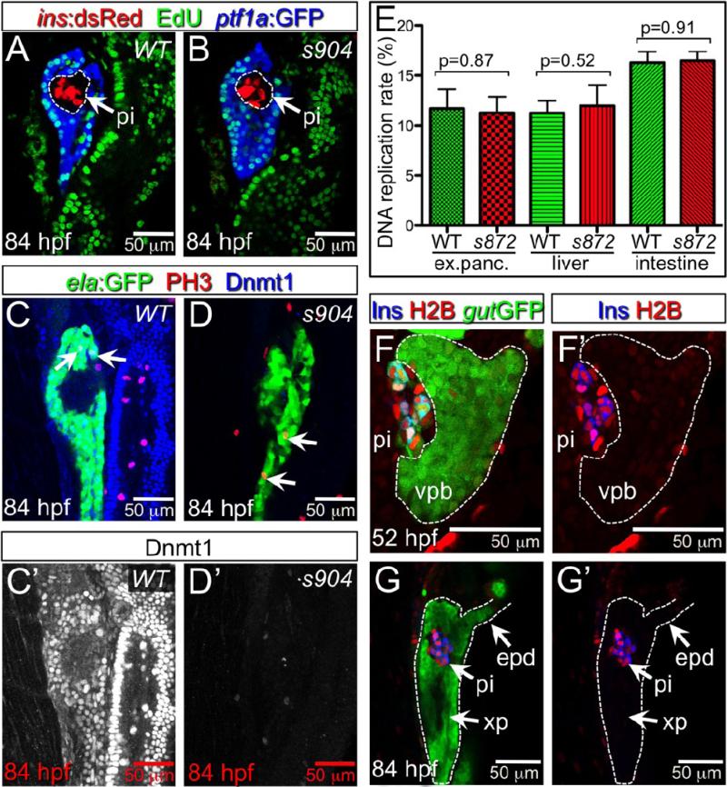
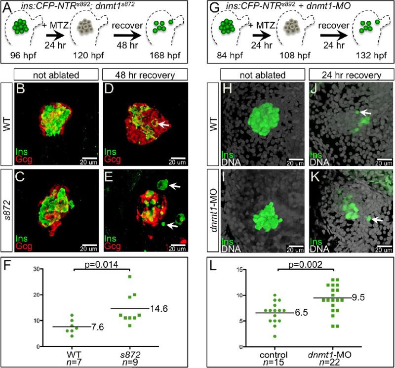
Developmental mechanisms regulating gene expression and the stable acquisition of cell fate direct cytodifferentiation during organogenesis. Moreover, it is likely that such mechanisms could be exploited to repair or regenerate damaged organs. DNA methyltransferases (Dnmts) are enzymes critical for epigenetic regulation, and are used in concert with histone methylation and acetylation to regulate gene expression and maintain genomic integrity and chromosome structure. We carried out two forward genetic screens for regulators of endodermal organ development. In the first, we screened for altered morphology of developing digestive organs, while in the second we screed for the lack of terminally differentiated cell types in the pancreas and liver. From these screens, we identified two mutant alleles of zebrafish dnmt1. Both lesions are predicted to eliminate dnmt1 function; one is a missense mutation in the catalytic domain and the other is a nonsense mutation that eliminates the catalytic domain. In zebrafish dnmt1 mutants, the pancreas and liver form normally, but begin to degenerate after 84 h post fertilization (hpf). Acinar cells are nearly abolished through apoptosis by 100 hpf, though neither DNA replication, nor entry into mitosis is halted in the absence of detectable Dnmt1. However, endocrine cells and ducts are largely spared. Surprisingly, dnmt1 mutants and dnmt1 morpholino-injected larvae show increased capacity for pancreatic beta cell regeneration in an inducible model of pancreatic beta cell ablation. Thus, our data suggest that Dnmt1 is dispensable for pancreatic duct or endocrine cell formation, but not for acinar cell survival. In addition, Dnmt1 may influence the differentiation of pancreatic beta cell progenitors or the reprogramming of cells toward the pancreatic beta cell fate.


Figures

Figure 1
Figure 1. Initial formation and degeneration of the exocrine pancreas in dandelion (ddn) mutants
(A-C) Lateral views of WT (A), ddns872 (B), and ddns904 mutant (C) larvae at 100 hpf in the 2-color Liver, Insulin, acinar Pancreas (2CLIP) transgenic background (see methods). Acinar cells of the exocrine pancreas (xp) are green and hepatocytes (li) and pancreatic beta cells (b) are red. ddn mutants exhibit minimal acinar tissue, but overall larva morphology is only moderately affected. (D,E) Successive images of individual WT (D) and ddns872 mutant (E) larvae between 84 and 148 hpf in the 2CLIP; Tg(ptf1a:EGFP)jh1 background. The exocrine pancreas and liver grow larger in WT, but degenerate in ddn mutants. Hepatocyte fragments are found in the circulation (arrowheads). (F-K) 3-D projections of confocal stacks showing composition of WT (F,H,J) and ddns872 mutant (G,I,K) endocrine pancreas at 100 hpf in Tg(ins:dsRed)m1081 background. (F,G) A core of Tg(ins:dsRed)m1081+ cells (red) is surrounded by a mantle of Glucagon+ (Gcg) cells (green). Extrapancreatic duct (epd) structure is intact. (H,I) Isl-1, and (J,K) Somatostatin (Sst) positive cells appear unaffected. Arrowheads in G, I, and M point to endocrine cells outside of the primary islet. (L,M) Nkx6.1 immunostaining reveals the presence of intrapancreatic duct cells in both WT and dnmt1s872 mutant larvae.
Figure 2
Figure 2. Liver degeneration in dandelion (ddn) mutants
(A-D) Lateral aspect of trunk (A,C) and tail (B,D) regions of 100 hpf WT (A,B) and ddn mutant (C,D) larvae in the 2CLIP transgenic background. The liver is smaller in ddn mutants relative to WT, and cell fragments emitting dsRed fluorescence, derived from the liver, aggregate in clusters in the tail (D, arrows). (E,F) 3-D projections of confocal stacks showing Alcam and Prox1 antibody staining of 100 hpf WT (E) and ddn mutants (F). Initial specification, morphogenesis, and differentiation of the liver appear to be unaffected. Intrahepatic ductal network is indicated by arrows. (G-J) Confocal slices through the liver (G,I) and tail vasculature (H,J) of Tg(kdrl:GFP)s843; Tg(fabp10:dsRed)gz12 WT (G,H) and ddn mutant (I,J) larvae at 6 dpf. Although never present in WT, bright dsRed+ degenerating hepatocyte fragments are observed throughout the ddn mutant liver (I, arrows) and within the hepatic vasculature (I, arrowheads). These particles accumulate in the network of the caudal vein (cv; J, arrows). Other abbreviations: ca, caudal aorta; se, intersegmental vessel; ib, intestinal bulb; p, pancreas; li, liver; b, beta cells.
Figure 3
Figure 3. The dandelion phenotype is caused by mutations in dnmt1
(A) Genetic map of the ddn region on linkage group 3; the numbers above the SSLP/RFLP markers indicate the number of recombinants in 932 meioses analyzed. Six genes lie within the critical region (shaded). (B) Sequence traces of pooled phenotypically WT and pooled dnmt1s872 mutant cDNAs; a G to A transversion at nucleotide 4376 results in a G1459D substitution in Dnmt1. (C) Sequence traces of pooled phenotypically WT and pooled dnmt1s904 mutant genomic DNA; a mutation in the exon 15 splice acceptor results in a single base pair frameshift and premature termination following 31 mis-sense substitutions. (D) Peptide sequence alignment of D. rerio (Dr) Dnmt1 from dnmt1s872 mutant and WT with H. sapiens (Hs), M. musculus (Mm), X. laevis (Xl), and A. thaliana (At). (E) Domain map of WT and ddn alleles of Dnmt1. (F) Southern blot of WT and dnmt1s872 and dnmt1s904 mutant genomic DNA either uncut or digested with methylation-sensitive HpaII or methylation insensitive MspI, and hybridized with a radiolabeled probe to the DANA consensus sequence. Appearance of DANA band (arrow) in dnmt1 mutant DNA digested with HpaII indicates hypomethylation of this transposable element, while smearing of high molecular weight DNA indicates widespread genomic hypomethylation (compare bracketed regions). (G-I) Dnmt1 antibody staining in 2CLIP transgenic line. Dnmt1 (blue) is observed in endodermal organs of WT (G) and dnmt1s872 mutants (H), but not dnmt1s904 mutants (I). (J-L) Injection of 4ng of dnmt1 morpholino (d1MO) into the 2CLIP line phenocopies ddn mutants. Compared to WT (J), d1MO-injected larvae (K) show smaller mass of differentiated pancreatic acinar cells (green) and hepatocytes (red) at 96 hpf. Ectopic dsRed fluorescence is observed in the posterior vasculature of d1MO-injected larvae at 168 hpf (L).
Figure 4
Figure 4. Acinar cell death in dnmt1 mutants occurs in part by p53-dependent apoptosis
(A,B) DRAQ5 staining of DNA in 84 hpf WT (A) and dnmt1s872 mutant (B) larvae. Pyknotic nuclei (arrows) are frequently observed in the pancreas of dnmt1 mutants, but not WT. (C,D) Apoptosis detection by TUNEL labeling of 84 hpf WT (C) and dnmt1 mutant (D) larvae in Tg(ptf1a:EGFP)jh1; Tg(ins:dsRed)m1081 background. C’/C’’ and D’/D’’ are transverse sections of similar samples at the approximate planes indicated in panels C and D, respectively. (E) Quantification of TUNEL+ acinar cells. (F-I) Expression of p53 mRNA in WT (F,H) and dnmt1s872 mutants (G,I); expression appears dramatically increased in mutants. (J) Fold-change of p53 and target genes mdm2 and p21/waf1 in dnmt1s872 mutants vs. WT by real-time RT-PCR. (K-N) 100 hpf WT (K,L) and dnmt1s872 mutant (M,N) larvae, which are either uninjected controls (K,M) or p53 morpholino-injected (p53MO; L,N). p53MO-injected dnmt1s872 mutants show increased persistence of acinar tissue. (O) More than 82% of p53MO-injected dnmt1s872 mutants showed rescue (green); n=22 (control), n=45 (p53MO). Error bars = SEM. Significance assessed by student's t-test.
Figure 5
Figure 5. Lack of Dnmt1 activity does not arrest the cell cycle
(A,B) Confocal planes showing EdU incorporation in 84 hpf WT (A) and dnmt1 mutant (B) Tg(ptf1a:EGFP)jh1; Tg(ins:dsRed)m1081 larvae. (C,D) Phospho-histone 3 (late G2 interphase through anaphase marker) and Dnmt1 staining of 84 hpf WT and dnmt1s904 mutant Tg(fabp10:DsRed; ela:GFP)gz12 larvae. (C’,D’) Dnmt1 channel only. (E) EdU incorporation rate in pancreatic acinar cells, hepatocytes, and intestine of dnmt1 mutants appears indistinguishable from WT (p=0.87, p=0.52, p=0.91, respectively). Error bars = SEM. Significance assessed by student's t-test. (F,G) H2BRFP label retention analysis of endodermal organ forming region in WT Tg(gut:GFP)s854 animals at 52 (F) and 84 (G) hpf. The ventral pancreatic bud (vpb) is demarcated by a dashed line. H2BRFP is diminished in the highly proliferative vpb relative to the dorsal pancreatic bud during outgrowth (F) and pancreas tail formation (G). (F’,G’) H2BRFP (red) and Ins (blue) channels only. Primary islet (pi).
Figure 6
Figure 6. Enhanced recovery of beta cell mass in dnmt1 mutants and morphants
(A) Pancreatic beta cell ablation scheme in dnmt1s872 mutants. 96 hpf Tg(ins:CFP-NTR)s892 WT and dnmt1 mutant larvae were exposed to MTZ for 24 hr, then washed for 48 hr and analyzed at 168 hpf. (B-E) Three-dimensional projections of the primary islet at 168 dpf in WT (B,D) and dnmt1 mutant (C,E) larvae that were untreated (B,C), or ablated with MTZ (D,E). (B,C) WT and dnmt1 mutant islets have a core of Ins+ beta cells surrounded by a mantle of Gcg+ alpha cells. (D) In recovering WT larvae, new beta cells are observed in the islet (arrow). (E) In recovering dnmt1 mutant islets, nearly double the number of beta cells are present, though some appear morphologically abnormal (arrows; compare with panel D). (F) 7.6 ± 1.0 beta cells were observed in recovering WT islets (n=7), and 14.6 ± 2.0 beta cells were observed in recovering dnmt1 mutant larvae (n=9; p=0.014). (G) Pancreatic beta cell ablation scheme in Tg(ins:CFPNTR)s892 embryos injected with 2 ng of dnmt1 morpholino (dnmt1-MO). 84 hpf larvae were exposed to MTZ for 24 hr, then washed for 24 hr and analysed at 132 hpf. (H-K) Three-dimensional projections of the primary islet at 132 hpf in WT (H,J) and dnmt1-MO injected (I,K) larvae that were untreated (H,I), or ablated with MTZ (J,K). (K) dnmt1-MO injected larvae showed a greater degree of beta cell recovery. (L) 6.5 ± 0.5 beta cells were observed in recovering WT larvae (n=15), and 9.5 ± 0.6 beta cells were observed in recovering dnmt1-MO injected larvae (n=22; p=0.002). Error bars = SEM. Significance assessed by student's t-test.

Similar articles

Cited by

References

    1. Beis D, Bartman T, Jin SW, Scott IC, D'Amico LA, Ober EA, Verkade H, Frantsve J, Field HA, Wehman A, Baier H, Tallafuss A, Bally-Cuif L, Chen JN, Stainier DY, Jungblut B. Genetic and cellular analyses of zebrafish atrioventricular cushion and valve development. Development. 2005;132:4193–204. - PubMed
    1. Biemar F, Argenton F, Schmidtke R, Epperlein S, Peers B, Driever W. Pancreas development in zebrafish: early dispersed appearance of endocrine hormone expressing cells and their convergence to form the definitive islet. Dev Biol. 2001;230:189–203. - PubMed
    1. Bird AP, Wolffe AP. Methylation-induced repression--belts, braces, and chromatin. Cell. 1999;99:451–4. - PubMed
    1. Blelloch R, Wang Z, Meissner A, Pollard S, Smith A, Jaenisch R. Reprogramming efficiency following somatic cell nuclear transfer is influenced by the differentiation and methylation state of the donor nucleus. Stem Cells. 2006;24:2007–13. - PMC - PubMed
    1. Bostick M, Kim JK, Esteve PO, Clark A, Pradhan S, Jacobsen SE. UHRF1 plays a role in maintaining DNA methylation in mammalian cells. Science. 2007;317:1760–4. - PubMed

Publication types

MeSH terms

Substances