

Functional connectivity and alterations in baseline brain state in humans
- PMID: 19631277
- PMCID: PMC2764802
- DOI: 10.1016/j.neuroimage.2009.07.028
Functional connectivity and alterations in baseline brain state in humans
Abstract
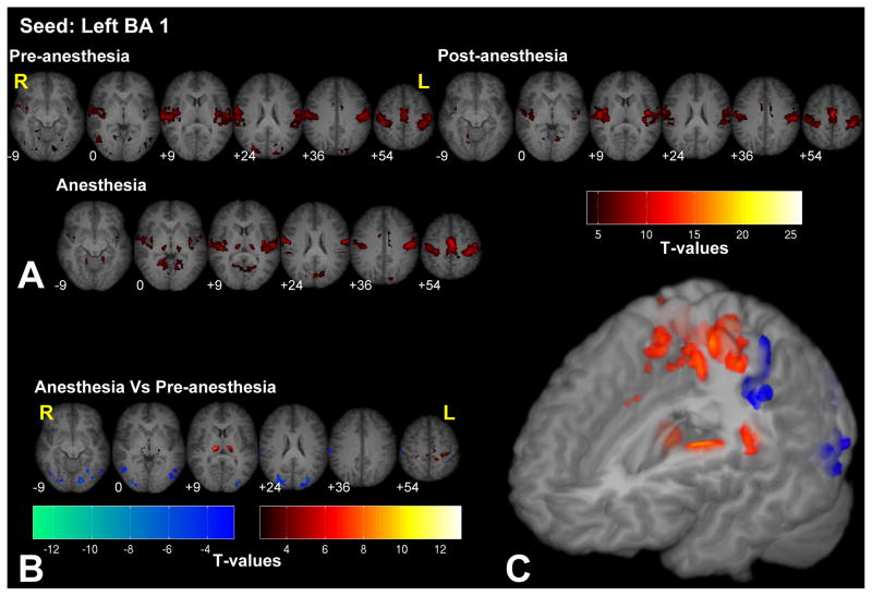
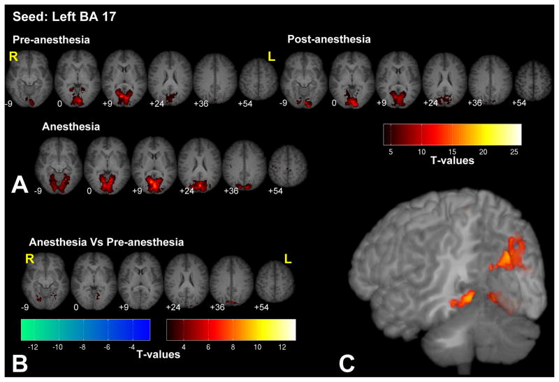
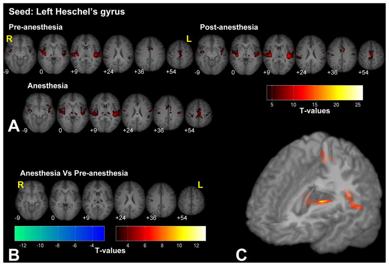
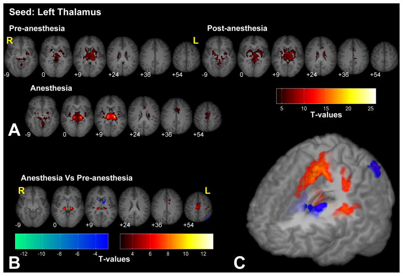
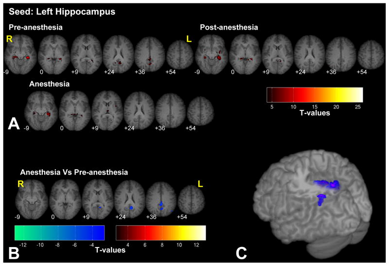
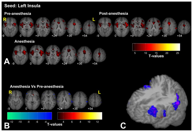
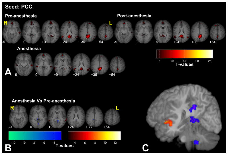
This work examines the influence of changes in baseline activity on the intrinsic functional connectivity fMRI (fc-fMRI) in humans. Baseline brain activity was altered by inducing anesthesia (sevoflurane end-tidal concentration 1%) in human volunteers and fc-fMRI maps between the pre-anesthetized and anesthetized conditions were compared across different brain networks. We particularly focused on low-level sensory areas (primary somatosensory, visual, and auditory cortices), the thalamus, and pain (insula), memory (hippocampus) circuits, and the default mode network (DMN), the latter three to examine higher-order brain regions. The results indicate that, while fc-fMRI patterns did not significantly differ (p<0.005; 20-voxel cluster threshold) in sensory cortex and in the DMN between the pre- and anesthetized conditions, fc-fMRI in high-order cognitive regions (i.e. memory and pain circuits) was significantly altered by anesthesia. These findings provide further evidence that fc-fMRI reflects intrinsic brain properties, while also demonstrating that 0.5 MAC sevoflurane anesthesia preferentially modulates higher-order connections.
Figures
Similar articles
-
Brain Resting-State Functional Connectivity Is Preserved Under Sevoflurane Anesthesia in Patients with Pervasive Developmental Disorders: A Pilot Study.Brain Connect. 2017 May;7(4):250-257. doi: 10.1089/brain.2016.0448. Brain Connect. 2017. PMID: 28443736
-
Altered local coherence in the default mode network due to sevoflurane anesthesia.Brain Res. 2010 Mar 8;1318:110-21. doi: 10.1016/j.brainres.2009.12.075. Epub 2010 Jan 6. Brain Res. 2010. PMID: 20059988 Free PMC article.
-
Sevoflurane 0.25 MAC preferentially affects higher order association areas: a functional magnetic resonance imaging study in volunteers.Anesth Analg. 2007 Sep;105(3):648-55. doi: 10.1213/01.ane.0000277496.12747.29. Anesth Analg. 2007. PMID: 17717218 Free PMC article. Clinical Trial.
-
Functional connectivity changes with concentration of sevoflurane anesthesia.Neuroreport. 2005 Feb 28;16(3):285-8. doi: 10.1097/00001756-200502280-00017. Neuroreport. 2005. PMID: 15706237 Clinical Trial.
-
The structural and functional connectivity of the posterior cingulate cortex: comparison between deterministic and probabilistic tractography for the investigation of structure-function relationships.Neuroimage. 2014 Nov 15;102 Pt 1:118-27. doi: 10.1016/j.neuroimage.2013.12.022. Epub 2013 Dec 21. Neuroimage. 2014. PMID: 24365673 Review.
Cited by
-
Intrinsic brain connectivity related to age in young and middle aged adults.PLoS One. 2012;7(9):e44067. doi: 10.1371/journal.pone.0044067. Epub 2012 Sep 11. PLoS One. 2012. PMID: 22984460 Free PMC article.
-
Global reduction of information exchange during anesthetic-induced unconsciousness.Brain Struct Funct. 2017 Sep;222(7):3205-3216. doi: 10.1007/s00429-017-1396-0. Epub 2017 Mar 13. Brain Struct Funct. 2017. PMID: 28289883 Free PMC article.
-
Low-motion fMRI data can be obtained in pediatric participants undergoing a 60-minute scan protocol.Sci Rep. 2020 Dec 14;10(1):21855. doi: 10.1038/s41598-020-78885-z. Sci Rep. 2020. PMID: 33318557 Free PMC article. Clinical Trial.
-
Propofol disrupts functional interactions between sensory and high-order processing of auditory verbal memory.Hum Brain Mapp. 2012 Oct;33(10):2487-98. doi: 10.1002/hbm.21385. Epub 2011 Sep 20. Hum Brain Mapp. 2012. PMID: 21932265 Free PMC article.
-
Sleep State Modulates Resting-State Functional Connectivity in Neonates.Front Neurosci. 2020 Apr 17;14:347. doi: 10.3389/fnins.2020.00347. eCollection 2020. Front Neurosci. 2020. PMID: 32362811 Free PMC article.
References
-
- Alkire MT, Haier RJ, Fallon JH. Toward a Unified Theory of Narcosis: Brain Imaging Evidence for a Thalamocortical Switch as the Neurophysiologic Basis of Anesthetic-Induced Unconsciousness. Conscious Cogn. 2000;9:370–386. - PubMed
-
- Alkire MT, Miller J, Steven L. Progress in Brain Research. Elsevier; 2005. General anesthesia and the neural correlates of consciousness; pp. 229–244.pp. 596–597. - PubMed
-
- Antognini JF, Buonocore MH, Disbrow EA, Carstens E. Isoflurane anesthesia blunts cerebral responses to noxious and innocuous stimuli: a fMRI study. Life Sci. 1997;61:PL349–PL354. - PubMed
-
- ASA Task Force. Practice guidelines for preoperative fasting and the use of pharmacologic agents to reduce the risk of pulmonary aspiration: application to healthy patients undergoing elective procedures: a report by the American Society of Anesthesiologist Task Force on Preoperative Fasting. Anesthesiology. 1999;90:896–905. - PubMed
Publication types
MeSH terms
Substances
Grants and funding
LinkOut - more resources
Full Text Sources
