Case Reports
. 2009 Aug;85(2):155-67.
doi: 10.1016/j.ajhg.2009.06.015. Epub 2009 Jul 23.

Identification of an agrin mutation that causes congenital myasthenia and affects synapse function

Affiliations
Case Reports

Identification of an agrin mutation that causes congenital myasthenia and affects synapse function

Caroline Huzé et al. Am J Hum Genet. 2009 Aug.

Erratum in

  • Am J Hum Genet. 2009 Oct;85(4):536

Abstract

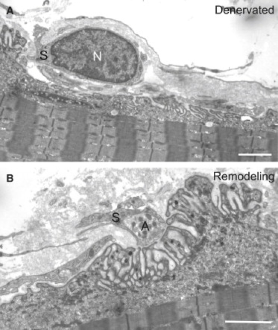
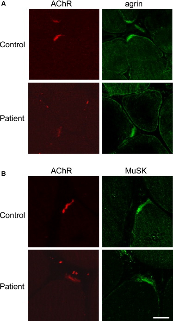
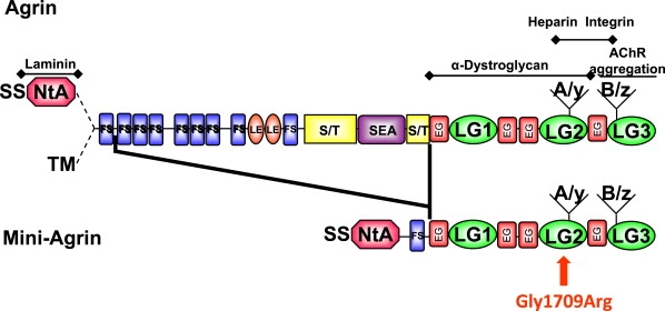
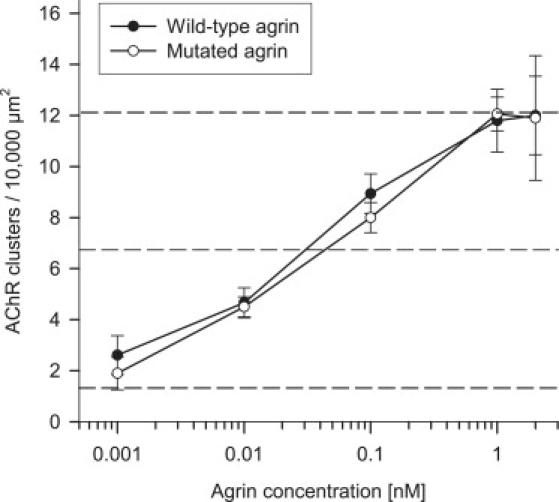
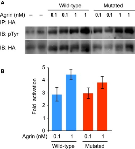
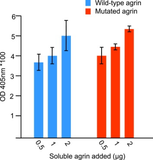
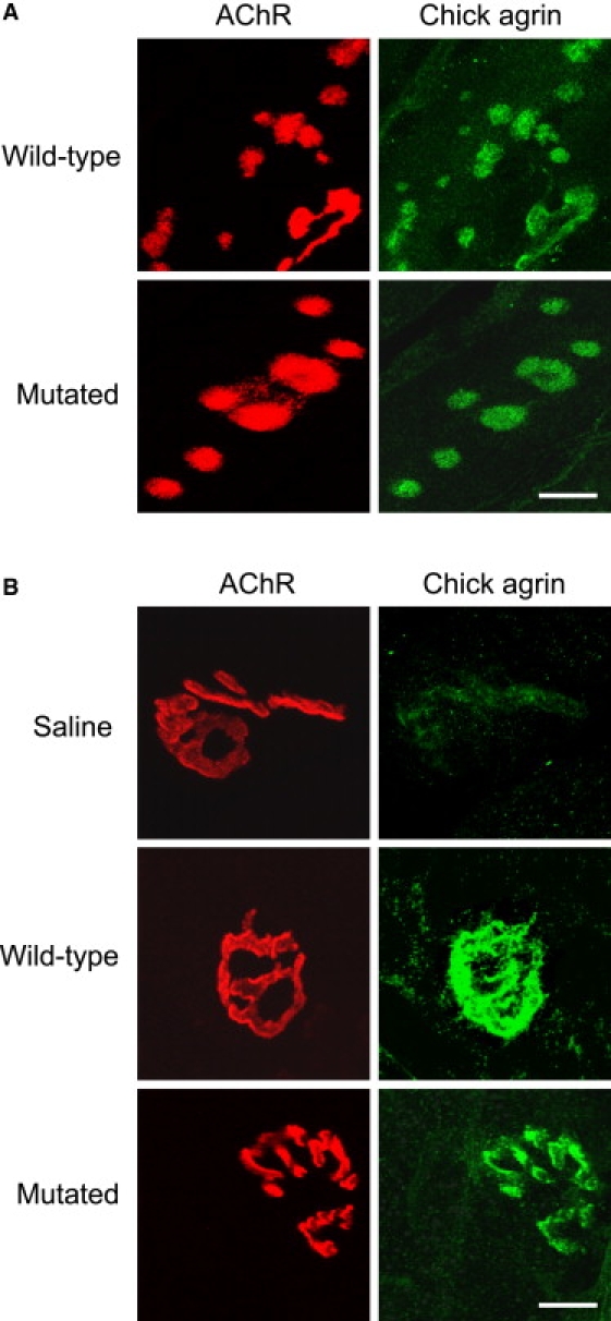
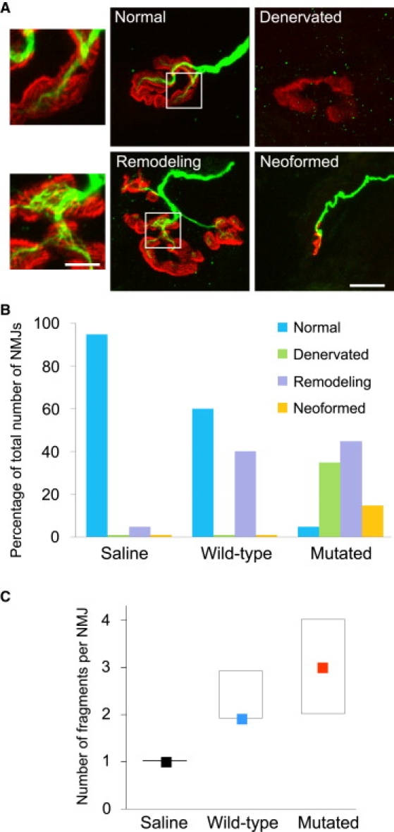
We report the case of a congenital myasthenic syndrome due to a mutation in AGRN, the gene encoding agrin, an extracellular matrix molecule released by the nerve and critical for formation of the neuromuscular junction. Gene analysis identified a homozygous missense mutation, c.5125G>C, leading to the p.Gly1709Arg variant. The muscle-biopsy specimen showed a major disorganization of the neuromuscular junction, including changes in the nerve-terminal cytoskeleton and fragmentation of the synaptic gutters. Experiments performed in nonmuscle cells or in cultured C2C12 myotubes and using recombinant mini-agrin for the mutated and the wild-type forms showed that the mutated form did not impair the activation of MuSK or change the total number of induced acetylcholine receptor aggregates. A solid-phase assay using the dystrophin glycoprotein complex showed that the mutation did not affect the binding of agrin to alpha-dystroglycan. Injection of wild-type or mutated agrin into rat soleus muscle induced the formation of nonsynaptic acetylcholine receptor clusters, but the mutant protein specifically destabilized the endogenous neuromuscular junctions. Importantly, the changes observed in rat muscle injected with mutant agrin recapitulated the pre- and post-synaptic modifications observed in the patient. These results indicate that the mutation does not interfere with the ability of agrin to induce postsynaptic structures but that it dramatically perturbs the maintenance of the neuromuscular junction.


Figures

Figure 1
Figure 1
Identification of the Mutation c.5125G>C in AGRN exon 29 and Hereditary Transmission (A) Sequence chromatograms from normal (wild-type) and affected (proband III-5) individuals are shown. (B) Family pedigree. The proband is indicated by an arrow. The expected nucleotide change p.Gly1709Arg transmitted in this consanguineous family is indicated below the symbols. ND: not determined.
Figure 2
Figure 2
Abnormal NMJs Observed in Patient Muscle-Biopsy Specimen (A) Whole-mount preparations stained for AChR with α-bungarotoxin in red and stained for axon terminals with neurofilament antibody in green. The different types of NMJs can be classified into four categories: normal, denervated, remodeling, and neoformed. The axonal branch classically ends as a fork and innervates a well-defined synaptic structure (normal). No axon profile faces the fragmented postsynaptic compartment (denervated). A thin axon terminal reinnervates some but not all of the fragmented gutters of the NMJ (remodeling). A thin axon innervating newly formed small and simplified NMJs can be observed (neoformed). In the bottom insert, note the disheveled appearance of the neurofilament staining observed in the NMJ in remodeling. The scale bar represents 10 μm for the four low-magnification prints and 4 μm for the two inserts. (B) The graph represents the classification of the NMJs observed in the patient muscle-biopsy specimen into four categories (expressed as a percentage of the 21 NMJs examined). (C) The fragmentation of NMJs was significantly increased in the patient in comparison with control muscle biopsies, according to the Mann-Whitney test (control: n = 28; patient: n = 21; U = 48,500, p < 0.001). Colored squares represent medians, and boxes represent 25th to 75th percentiles.
Figure 3
Figure 3
Electron Micrographs of Two NMJs from the Patient Biopsy Specimen (A) An emptied NMJ is fully engulfed by a terminal Schwann cell (S) whose nucleus is clearly visible (N). (B) A small reinnervating axon terminal (A) partially encased by Schwann cell processes (S) intrudes into the synaptic cleft, and there is pronounced reduction of the area of apposition between the axon terminal (A) and the postsynaptic membrane. The scale bar represents 2 μm in both prints.
Figure 4
Figure 4
Cross-Sections of the Patient Biopsy Specimen Were Double Labeled for AChR in Red and for Agrin or MuSK in Green Both agrin (A) and MuSK (B) colocalize with AChR at the NMJ, and their relative expression between the control and patient appears roughly similar. The scale bar represents 20 μm and applies to the eight prints.
Figure 5
Figure 5
Protein Domains of the Recombinant Mini-Agrin and Localization of the p.Gly1709Arg Substitution The neural mini-agrin is deleted from its central region. It contains the SS-NtA domain at the N-terminal region and the A/y and B/z inserts at the C-terminal region. The p.Gly1709Arg substitution is located in the laminin G-like 2 domain (LG2) in the region of agrin interaction with α-dystroglycan, heparin, and integrins.
Figure 6
Figure 6
Effects of the Mutated Agrin on AChR Aggregation Dose-response curves for AChR-aggregating activity of wild-type or mutant agrins. C2C12 myotubes were incubated for 18 hr with recombinant agrins at the concentrations indicated, and the number of AChR clusters per myotube was determined as described in the Material and Methods. Each data point represents results (mean ± SD) from triplicate myotube cultures; ten fields were studied per dish. Data shown represent the results of one representative experiment. The number of AChR clusters without agrin and the half-maximal and maximal number of clusters are indicated by dotted lines.
Figure 7
Figure 7
Effects of the Mutated Agrin on MuSK Phosphorylation 293T cells expressing both MuSK and LRP4 were incubated with wild-type and mutant agrin at different concentrations (0.1–5 nM). A representative experiment is shown with 0.1 and 1 nM agrin. (A) HA-MuSK was immunoprecipitated (IP) with an anti-HA antibody (HA), and immunoblots (IB) for MuSK (HA) and phosphorylated MuSK (pTyr) were performed. (B) Phosphorylation, estimated with ImageJ software, is expressed as n-fold activation. No significant differences between wild-type or mutant agrin were observed at any concentration studied.
Figure 8
Figure 8
Evaluation of Agrin Binding to DGC in Solid-Phase Assay Various amounts of wild-type and mutant agrin were added to 0.5 μg of DGC bound to 96-well plates. The amount of soluble agrin added is indicated in micrograms on the abscissa. The amount of bound agrin is expressed as absorbance ± SD.
Figure 9
Figure 9
Chick Agrin Colocalizes with Extrasynaptic AChR Clusters and Endogenous NMJs in Both Wild-Type and Mutated Agrin-Injected Muscle (A) Agrin-induced extrasynaptic clusters are double labeled for AChR in red and for chick agrin in green and observed by confocal microscopy. The calibration bar represents 4 μm. (B) Native NMJs are double labeled for AChR in red and for chick agrin in green and observed by confocal microscopy. The calibration bar represents 10 μm. Note that chick agrin is present at NMJs in both wild-type and mutated-agrin-injected muscle, but not in saline-injected rat soleus muscle.
Figure 10
Figure 10
Mutated Agrin Induces Remodeling of Pre-existing NMJs and Their Fragmentation (A) Rat soleus muscles were injected with saline, wild-type agrin, or mutated agrin. After 2 weeks, muscles were removed and stained for AChRs with α-bungarotoxin (red) and neurofilament antibodies (green). As for the patient biopsy specimen (see Figure 2), the different NMJs observed were divided into four categories: normal, denervated, remodeling, and neoformed. Note in the bottom insert the disheveled appearance of the stained neurofilaments observed in the NMJ in remodeling. This peculiar aspect of disassembly of terminal axons was specific of NMJs after injection of mutated agrin and was never observed after injection of wild-type agrin. The scale bar represents 10 μm for the four low-magnification prints and 4 μm for the two inserts. (B) The graph represents the distribution of the NMJs in these four categories depending on the injection: saline, wild-type agrin, or mutated agrin. Note that the distribution for mutated agrin is similar to that observed in the patient muscle-biopsy specimen (Figure 2B). (C) The fragmentation of NMJs was significantly increased in mutated agrin-treated muscles in comparison with the saline-treated and even the wild-type agrin-treated muscles according to Kruskal-Wallis test (saline: n = 42; wild-type agrin: n = 45; mutated agrin: n = 44; H = 52; p < 0.001). Colored squares represent medians, and boxes represent 25th to 75th percentiles.

References

    1. Hantaï D., Richard P., Koenig J., Eymard B. Congenital myasthenic syndromes. Curr. Opin. Neurol. 2004;17:539–551. - PubMed
    1. Engel A.G., Sine S.M. Current understanding of congenital myasthenic syndromes. Curr. Opin. Pharmacol. 2005;5:308–321. - PubMed
    1. Müller J.S., Mihaylova V., Abicht A., Lochmüller H. Congenital myasthenic syndromes: Spotlight on genetic defects of neuromuscular transmission. Expert Rev. Mol. Med. 2007;9:1–20. - PubMed
    1. Maselli R.A., Ng J.J., Anderson J.A., Cagney O., Arredondo J., Williams C., Wessel H.B., Abdel-Hamid H., Wollmann R.L. Mutations in LAMB2 causing a severe form of synaptic congenital myasthenic syndrome. J. Med. Genet. 2009;46:203–208. - PMC - PubMed
    1. Chevessier F., Faraut B., Ravel-Chapuis A., Richard P., Gaudon K., Bauché S., Prioleau C., Herbst R., Goillot E., Ioos C. MUSK, a new target for mutations causing congenital myasthenic syndrome. Hum. Mol. Genet. 2004;13:3229–3240. - PubMed

Publication types

MeSH terms