. 2009 Aug 7;138(3):576-91.
doi: 10.1016/j.cell.2009.06.015. Epub 2009 Jul 23.

RNA polymerase III detects cytosolic DNA and induces type I interferons through the RIG-I pathway

Affiliations

RNA polymerase III detects cytosolic DNA and induces type I interferons through the RIG-I pathway

Yu-Hsin Chiu et al. Cell. .

Abstract

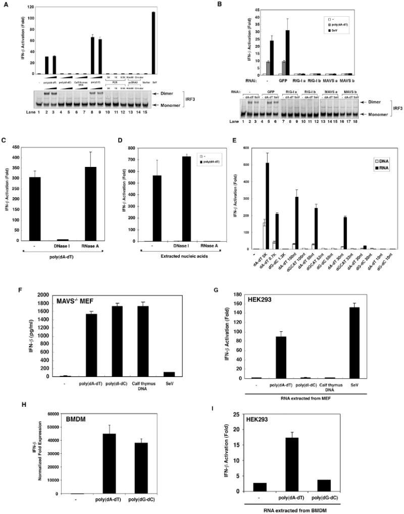
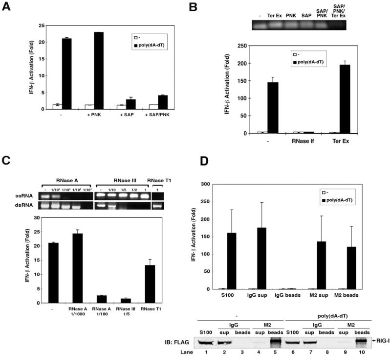
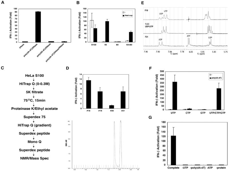
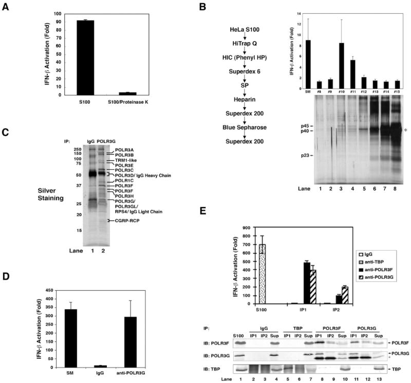
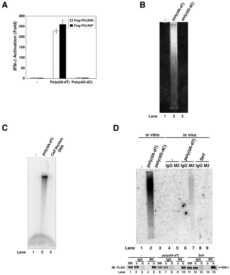
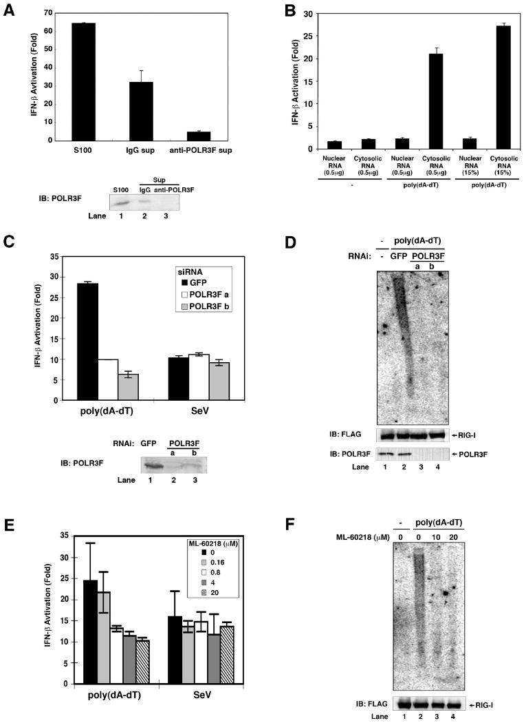
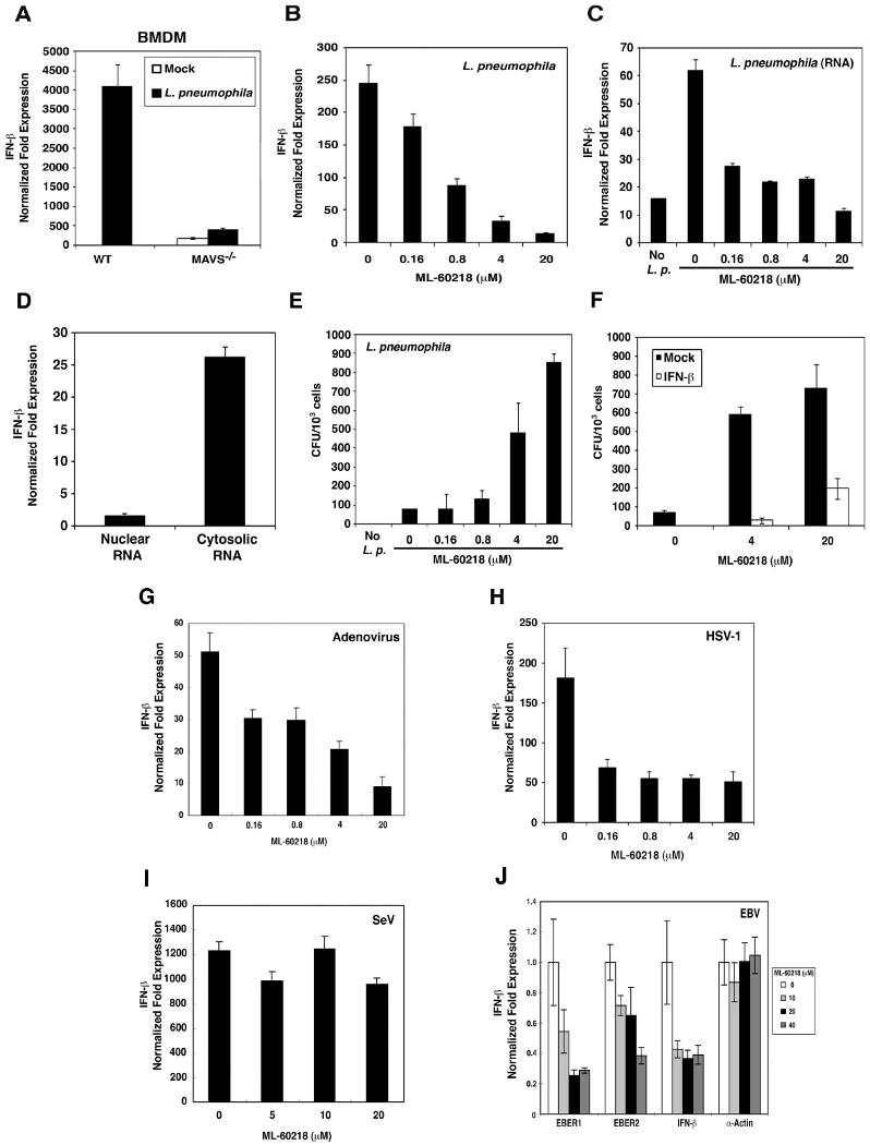
Type I interferons (IFNs) are important for antiviral and autoimmune responses. Retinoic acid-induced gene I (RIG-I) and mitochondrial antiviral signaling (MAVS) proteins mediate IFN production in response to cytosolic double-stranded RNA or single-stranded RNA containing 5'-triphosphate (5'-ppp). Cytosolic B form double-stranded DNA, such as poly(dA-dT)*poly(dA-dT) [poly(dA-dT)], can also induce IFN-beta, but the underlying mechanism is unknown. Here, we show that the cytosolic poly(dA-dT) DNA is converted into 5'-ppp RNA to induce IFN-beta through the RIG-I pathway. Biochemical purification led to the identification of DNA-dependent RNA polymerase III (Pol-III) as the enzyme responsible for synthesizing 5'-ppp RNA from the poly(dA-dT) template. Inhibition of RNA Pol-III prevents IFN-beta induction by transfection of DNA or infection with DNA viruses. Furthermore, Pol-III inhibition abrogates IFN-beta induction by the intracellular bacterium Legionella pneumophila and promotes the bacterial growth. These results suggest that RNA Pol-III is a cytosolic DNA sensor involved in innate immune responses.


Figures

Figure 1
Figure 1. Poly(dA-dT) activates RIG-I-dependent IFN-β production through intermediary RNA
(A) HEK293 cells were transfected with the reporter plasmid IFN-β-Luc (25 ng/ml), pCMV-LacZ (50 ng/ml) and various types of DNA or RNA [poly(I:C)] as indicated (1 μg/ml). Cells were lysed 16 hours after transfection, followed by luciferase reporter assay (upper panel) or IRF3 dimerization assay (lower panel). PCR: linear PCR fragments of the indicated size; HindIII: HindIII-linearized fragment of pcDNA3; marker: DNA marker (0.1-10 kB; NEB); SeV: Sendai virus infection. (B) HEK293 cells were transfected with control siRNA oligos against GFP, two different pairs of siRNA oligos (a & b) against RIG-I, or MAVS. Subsequently, cells were cotransfected with IFN-β-Luc, pCMV-LacZ and pcDNA3. After 24 hours, cells were transfected with poly(dA-dT) (1 μg/ml) or infected with SeV. Luciferase reporter assay (upper panel) or IRF3 dimerization assay (lower panel) were performed 16 hours after transfection or infection. (C) HEK293 cells were transfected with poly(dA-dT) which had been pre-digested with DNase I (0.2 U/μl) or RNaseA (0.1 mg/ml), and then IFN-β luciferase reporter assay was carried out as described in (A). (D) HEK293 cells were transfected with or without poly(dA-dT) for 16 hours, and then nucleic acids were prepared from the cell lysates by phenol/chloroform extraction. After digestion with DNase I (0.2 U/μl) or RNase A (0.1 mg/ml), the nucleic acids were transfected into HEK293-IFNβ-luciferase reporter cells to measure IFN-β induction. (E) HEK293 cells were transfected with DNA of different sizes and compositions as indicated (1 μg/ml). Total RNA was extracted with TRIzol and then transfected into HEK293-IFNβ-luciferase reporter cells. In parallel experiments, the DNA was directly transfected into HEK293-IFNβ-luciferase reporter cells. (F) MAVS-deficient MEF cells were transfected with various DNA or infected with SeV. Culture supernatants were collected 16 hours after treatment for measurement of IFN-β by ELISA. (G) MAVS-deficient MEF cells were treated as described in (F) and then total RNAs were extracted by TRIzol and transfected into HEK293-IFNβ-luciferase reporter cells. (H) Mouse BMDM were transfected with poly(dA-dT) or poly(dG-dC) for 8 hours before total RNAs were extracted for qPCR. The expression level of IFN-β gene was normalized with that of β-actin gene. (I) BMDM cells were transfected as described in (H) and then total RNAs were extracted by TRIzol and transfected into HEK293-IFNβ-luciferase reporter cells.
Figure 2
Figure 2. Properties of the IFN-inducing RNA derived from poly(dA-dT)
(A) RNAs extracted from poly(dA-dT) or mock transfected cells were treated with shrimp alkaline phosphatases (SAP) or polynucleotide kinase (PNK) at 37°C for 1 hour. An aliquot of SAP-treated RNA was further treated with PNK at 37°C for 1 hour to phosphorylate the DNA. RNAs were precipitated by ethanol precipitation and then transfected into HEK293-IFNβ-luciferase reporter cells. (B) Upper panel: poly(A-U) RNA transcribed by T7 polymerase was treated with terminator exonuclease (Ter Ex), PNK, SAP at 37°C for 1 hour. Two aliquots of SAP-treated RNA were phosphorylated with PNK before one of the aliquots was further treated with Ter Ex. Lower panel: RNAs extracted from poly(dA-dT)- or mock-transfected cells were treated with RNAse-If or Ter Ex and then transfected into HEK293-IFNβ-luciferase reporter cells. (C) Upper panels: single-stranded RNA (ssRNA) or double-strand RNA (dsRNA) generated by T7 polymerase was digested with various amounts of RNase A, RNase III, or RNase T1 as indicated, separated on 1% agarose gel and then visualized by ethidium bromide staining. Lower panel: RNAs extracted from poly(dA-dT) transfected cells were digested with RNAse A, RNase III, or RNase T1 and then transfected into HEK293-IFNβ-luciferase reporter cells. (D) Poly(dA-dT) was transfected into HEK293 cells stably expressing FLAG-tagged RIG-I, which was subsequently immunoprecipitated with an anti-FLAG antibody (M2) or control IgG. Nucleic acids in the cell extracts (S100), unbound supernatant (sup) and immunoprecipitates (beads) were extracted and then transfected into HEK293-IFNβ-luciferase reporter cells (upper panel). An aliquot of S100, sup and beads was separated by SDS-PAGE and immunoblotted with a FLAG antibody (lower panel).
Figure 3
Figure 3. In vitro generation of IFN-inducing RNA requires ATP and UTP
(A) HeLa S100 was incubated with poly(dA-dT) or poly(dG-dC) (20 μg/ml) and ATP (2mM) at 30°C for 1 hour. RNA was extracted with phenol/chloroform after DNase I (0.2 U/μl) and/or RNase A (0.1mg/ml) treatment, then transfected into HEK293-IFNβ-luciferase reporter cells. (B) HeLa S100 was precipitated with 40% ammonium sulfate, then the supernatant was further precipitated with 80% ammonium sulfate. The precipitates were dialyzed and incubated with poly(dA-dT) and ATP in the presence or absence of the supernatant from heat-treated HeLa S100 (“heat sup”). RNA from the in vitro reaction was extracted and transfected into HEK293-IFNβ-luciferase reporter cells. (C) Scheme of purification of heat-resistant factor required for the generation of IFN-inducing RNA. (D) Chromatogram (A260) of the small molecules on Superdex peptide column (last step; lower panel). Factions from the Superdex peptide column were incubated with a protein fraction from 40% ammonium sulfate precipitation, poly(dA-dT) and ATP, and then the RNA were extracted for IFN-β reporter assays (upper panel). (E) NMR spectra of fractions 18 (F18; top spectrum) and 21 (F21; bottom). The middle spectrum is a mixture of authentic UDP and UTP standards (1:2.5). (F) UTP, CTP, GTP or UTP/CTP/GTP (1.5 mM) was used to substitute for the “heat sup” in the in vitro reaction, and then RNA was extracted for IFN-β reporter assays. (G) Reactions containing a crude protein fraction (40% ammonium sulfate precipitate), poly(dA-dT), ATP and UTP (complete reaction) or lacking one of the components as indicated were carried out in vitro, and then RNA was extracted for IFN-β reporter assays.
Figure 4
Figure 4. Purification and identification of Pol-III
(A) HeLa S100 was incubated with or without proteinase K (2 mg/ml) at 37 °C for 1 hour and then incubated with poly(dA-dT) and ATP before RNA was extracted for IFN-β reporter assays. (B) Protein purification was carried out according to the scheme shown on the left and frations from the last Superdex 200 column were analyzed for their activity to produce IFN-inducing RNA (upper panel) and by silver staining (12% SDS-PAGE; lower panel). Arrows indicate the proteins co-purifying with the activity and analyzed by mass spectrometry. The asterisk indicates chicken albumin which was added as a carrier protein during purification. (C) Cytosolic extracts from HEK293 cells stably expressing FLAG-POLR3F were used for immunopurification of the Pol-III complex using anti-FLAG agarose (M2). Following elution with FLAG peptide, aliquots of the FLAG-Pol-III complex were subjected to immunoprecipitation with an antibody against POLR3G or control IgG, and the precipitated proteins were analyzed by silver staining. Unique bands from anti-POLR3G were identified by mass spectrometry. (D) The immunopurified Pol-III complex from (C) was incubated with poly(dA-dT), ATP and UTP before RNA was extracted for IFN-β reporter assays. (E) HeLa cell lysate (S100) was immunoprecipitated twice (IP1 and IP2) with an antibody specific for TBP, POLR3F, POLR3G or control IgG. The precipitated endogenous proteins were tested for their activity to produce IFN-inducing RNA (upper panel). Aliquots of the precipitated proteins and supernatants (“Sup”) were analyzed by immunoblotting with the indicated antibodies.
Figure 5
Figure 5. Pol-III catalyzes the synthesis of poly(A-U) RNA using poly(dA-dT) as the template
(A) Pol-III complex was immunopurified from HEK293 cells stably expressing FLAG-tagged POLR3D or POLR3F, and then incubated with poly(dA-dT) or poly(dG-dC) in the presence of NTP. RNA was extracted from the reactions for IFN-β reporter assays. (B) RNAs synthesized by in vitro transcription of poly(dA-dT) or poly(dG-dC) were analyzed by agarose gel eletrophoresis followed by ethidium bromide staining. (C) RNAs synthesized by in vitro transcription of poly(dA-dT) or calf thymus DNA in the presence of α-32P-UTP and NTP were analyzed by agarose gel electrophoresis followed by autoradiography. (D) HEK293 cells stably expressing FLAG-tagged RIG-I were transfected with poly(dA-dT) (lanes 6 & 7), infected with Sendai virus (lanes 8 & 9) or mock treated (lanes 4 & 5). RIG-I was subsequently immunoprecipitated with the FLAG antibody (M2) or control IgG, and RNA in the precipitates was extracted and analyzed by Northern blotting using 32P-labeled poly(A-U) RNA as a probe. In lanes 1-3, RNAs synthesized by in vitro transcription of poly(dA-dT) or poly(dG-dC) or in the absence of DNA were analyzed by Northern blotting. Bottom panel: Immunoprecipitated proteins were analyzed by immunoblotting with a FLAG antibody. SM: starting material (cell lysates); s: supernatant; b: bound proteins on the beads.
Figure 6
Figure 6. Pol-III is required for the production of IFN-inducing RNA
(A) HeLa cell lysate (S100) was immunodepleted five times with a POLR3F specific antibody or control IgG. S100 and the immunodepleted supernatants were incubated with poly(dA-dT), UTP and ATP before RNA was extracted for IFN-β reporter assays (upper panel). The efficiency of immunodepletion was analyzed by immunoblotting with the POLR3F antibody (lower panel). (B) HEK293 cells were transfected with or without poly(dA-dT) for 4 hours. Nuclear and cytosolic lysates were prepared for RNA extraction. 0.5 μg or 15% of nuclear or cytosolic RNAs were transfected into HEK293-IFNβ-luciferase reporter cells to measure IFN-β induction. (C) Two distinct siRNA oligos against POLR3F (a and b) or a control siRNA against GFP were transfected into HEK293 cells. 48 hours after transfection, the cells were transfected with poly(dA-dT) or infected with Sendai virus for 2 hours before RNAs were extracted for IFN-β reporter assays (upper). The efficiency of RNAi was analyzed by immunoblotting with an antibody against POLR3F (lower). (D) HEK293/RIG-I-FLAG stable cells were transfected with siRNA as described in (C), then transfected with poly(dA-dT) for 3 hours. The RIG-I complex was immunoprecipitated using a FLAG antibody, then the RNAs associated with RIG-I were extracted and analyzed by Northern blotting using 32P-labeled poly(A-U) RNA as a probe. The RIG-I in the immunoprecipitates and POLR3F in cell lysates were analyzed by immunoblotting. (E) HEK293 cells were treated with the indicated concentrations of the Pol-III inhibitor ML-60218 for 10 hours before transfection with poly(dA-dT) or infection with Sendai virus for 2 hours. RNAs were extracted for IFN-β reporter assays. (F) HEK293/RIG-I-FLAG stable cells were treated with ML-60218 and then transfected with poly(dA-dT). RNAs associated with the RIG-I complex were analyzed by Northern blotting as described in (D).
Figure 7
Figure 7. Pol-III is required for IFN-β induction by intracellular bacteria and DNA viruses
(A) Wild-type or MAVS-deficient BMDM cells were infected with Legionella pneumophila for 8 hours. Total RNAs were extracted for qPCR. The expression level of IFN-β gene was normalized with that of β-actin gene. (B) Raw264.7 cells were treated with the indicated concentrations of ML-60218 for 10 hours and then infected with Legionella pneumophila. Total RNAs were extracted after 8 hours of infection for qPCR of IFN-β and β-actin. (C) RNAs extracted from ML-60218-treated and Legionella pneumophila-infected Raw264.7 cells as described in (B) were transfected into Raw264.7 cells for 8 hours. The expression of IFN-β and β-actin was measured by qPCR. (D) Raw264.7 cells were infected with Legionella pneumophila for 8 hours. Nuclear and cytosolic lysates were prepared to extract RNAs, which were transfected into Raw264.7 cells for 8 hours. The expression of IFN-β was measured by qPCR and normalized to the level of β-actin. (E) Raw264.7 cells were treated with ML-60218 for 10 hours and infected with Legionella pneumophila for 24 hours. Cells were lysed in 0.1 % saponin and the lysates were spread on BCYE agar plates. Bacterial colonies were counted 2 days after incubation and shown as CFU per 1000 cells. (F) Raw264.7 cells were treated with ML-60218 in the presence or absence of mouse IFN-β (1000 U/ml) for 10 hours and subsequently infected with Legionella pneumophila for 24 hours. Bacterial colonies were determined as described in (E). (G-I) Raw264.7 cells were treated with ML-60218 for 10 hours and then infected with adenovirus (AdV; G), Herpes Simplex Virus-1 (HSV-1; H) or Sendai virus (SeV; I). Total RNAs were extracted for qPCR of IFN-β and β-actin. (J) The Epstein-Bar virus (EBV)-producing B95-8 cells were treated with ML-60218 for 24 hours and then total RNAs were prepared for qPCR. The expression levels of EBER1, EBER2, IFN-β and α-actin genes were normalized with that of the β-actin RNA. Both α- and β-actin are Pol-II transcribed genes.

Comment in

Similar articles

Cited by

References

    1. Akira S, Uematsu S, Takeuchi O. Pathogen recognition and innate immunity. Cell. 2006;124:783–801. - PubMed
    1. Cheng G, Zhong J, Chung J, Chisari FV. Double-stranded DNA and double-stranded RNA induce a common antiviral signaling pathway in human cells. Proc Natl Acad Sci U S A. 2007;104:9035–9040. - PMC - PubMed
    1. Deng L, Wang C, Spencer E, Yang L, Braun A, You J, Slaughter C, Pickart C, Chen ZJ. Activation of the IkappaB kinase complex by TRAF6 requires a dimeric ubiquitin-conjugating enzyme complex and a unique polyubiquitin chain. Cell. 2000;103:351–361. - PubMed
    1. Hornung V, Ellegast J, Kim S, Brzozka K, Jung A, Kato H, Poeck H, Akira S, Conzelmann KK, Schlee M, et al. 5′-Triphosphate RNA is the ligand for RIG-I. Science. 2006;314:994–997. - PubMed
    1. Howe JG, Shu MD. Upstream basal promoter element important for exclusive RNA polymerase III transcription of the EBER 2 gene. Mol Cell Biol. 1993;13:2655–2665. - PMC - PubMed

Publication types

MeSH terms