Review
. 2009 Aug;19(4):425-32.
doi: 10.1016/j.sbi.2009.06.002. Epub 2009 Jul 22.

Structure and function of Na(+)-symporters with inverted repeats

Affiliations
Review

Structure and function of Na(+)-symporters with inverted repeats

Jeff Abramson et al. Curr Opin Struct Biol. 2009 Aug.

Abstract

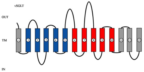
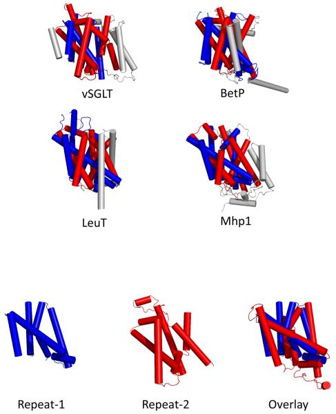
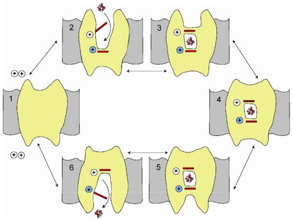
Symporters are membrane proteins that couple energy stored in electrochemical potential gradients to drive the cotransport of molecules and ions into cells. Traditionally, proteins are classified into gene families based on sequence homology and functional properties, for example the sodium glucose (SLC5 or Sodium Solute Symporter Family, SSS or SSF) and GABA (SLC6 or Neurotransmitter Sodium Symporter Family, NSS or SNF) symporter families [1-4]. Recently, it has been established that four Na(+)-symporter proteins with unrelated sequences have a common structural core containing an inverted repeat of 5 transmembrane (TM) helices [5(**)-8(**)]. Analysis of these four structures reveals that they reside in different conformations along the transport cycle providing atomic insight into the mechanism of sodium solute cotransport.


Figures

Figure 1
Figure 1. 2-D structure of vSGLT
A 2D model of vSGLT highlighting the core domain of 10 TMs composed of inverted repeats of 5TMs. vSGLT is a 543 amino acid protein containing 14 TMs with N- and C-termini on the extracellular face of the membrane [41,42]. To avoid confusion, we number the TMs in the inverted repeats as in LeuT ([5], TM1-TM5 (Blue) and TM6-TM10 (red). As members of families contain up to 15 TMs, addition N-terminal TMs are numbered TM(-1) (vSGLT), TM(-1) and TM(-2) (BetP) and additional C-terminal TMs are numbered TM11 and TM12 (LeuT, Mhp1) and TM11, TM12, and TM13 (vSGLT). Note: the N-terminus of the first repeat is intracellular while that for the inverted repeat is extracellular.
Figure 2
Figure 2. The 3D structures of the sodium symporters
A) Cylinder representation of vSGLT, BetP, LeuT and Mhp1 structures. All four structures have a core domain of 10 transmembrane (TM) helices formed from an internal structural repeat. B) The two structural repeats have an inverted topology where TM1-TM5 is related to TM6-TM10 by an apparent two-fold symmetry around an axis through the center of the membrane plane permitting superimposition. A superposition of vSGLT's TM1-TM5 and inverted TM6-TM10 yields an RMSD of 3.9 Å for 131 Cα atoms. TM1-TM5 and TM6-TM10 are colored red and blue respectively with additional helices on the N- and C-termini colored gray.
Figure 3
Figure 3. The location of substrate and the common sodium binding site
A) Structural alignment of the core domains from vSGLT, BetP, LeuT and Mhp1 yield an RMSD between 3.8 Å and 4.5 Å. TM1-TM5 and TM6-TM10 are colored red and blue respectively. B) Side view of vSGLT showing the location of the sugar binding site, the extracellular (M73, Y87 and F424) and intracellular (Y263) gates. The numbering of the TMs is according to the convention shown in Figure 1. C) A structural alignment of TM1, TM6 and the substrate-binding site for vSGLT (red), BetP (blue), LeuT (green) and Mhp1 (cyan). D) A structural alignment of TM1, TM8 and the conserved sodium-binding site for vSGLT (red), BetP (blue), LeuT (green) and Mhp1 (cyan).
Figure 4
Figure 4. A Simple 6-state model for sodium-substrate symport
In this model the starting point is state 1 (C1) with no ligands bound. In the presence of extracellular sodium, one (or two) Na+ ions bind first to open the external gates to the substrate binding site (C2). External substrate is then able to bind to its binding site and this in turn closes the external gate (C3). The next step is closure of the external vestibule (C4) and this is followed by opening of the internal vestibule (C5). Opening of the internal gate permits substrate and sodium ion(s) to dissociate and exit at the intracellular face of the membrane (C6). The cycle is completed by closure of the internal vestibule and the return to C1. The complete cycle results in the transport of sodium and substrate across the membrane in a fixed stoichiometry (1/1, 2/1). The direction of transport is determined by the ligand concentrations on each side of the membrane and the membrane potential [14,25,27,31-33,43]. The crystal structures of Mhp1 appear to correspond to C2 and C3, LeuT to C3, BetP to C4 and vSGLT to C5. At least in the case of vSGLT the monomer is completely functional [41].

Similar articles

Cited by

References

    1. Nelson N. The Family of Na+/C1- Neuortransmitter Transporters. Journal of Neurochemistry. 199871:1786–1803. - PubMed
    1. Wright EMaT E. The Sodium Glucose Cotransport Family SLC5. Pflugers Arch. 2004:510–518. - PubMed
    1. Turk E, Wright EM. Membrane topology motifs in the SGLT cotransporter family. J Membr Biol. 1997;159:1–20. - PubMed
    1. Chen NH, Reith ME, Quick MW. Synaptic uptake and beyond: the sodium-and chloride-dependent neurotransmitter transporter family SLC6. Pflugers Arch. 2004;447:519–531. - PubMed
    1. Yamashita A, Singh SK, Kawate T, Jin Y, Gouaux E. Crystal structure of a bacterial homologue of Na+/Cl--dependent neurotransmitter transporters. Nature. 2005;437:215–223. - PubMed
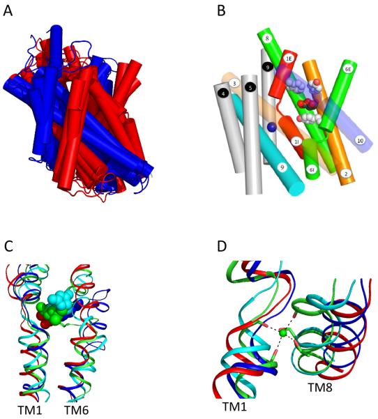
    2. The first high resolution crystal structure of a sodium symporter with the inverted 5TM motif containing a pair of transmembrane discontinuous helices. The substrate, leucine, and one sodium was bound in a occluded site halfway across the membrane close to these two discontinuous helices. A route for substrate entry to the binding site from the external solution was identified and this conformation of the protein was termed the outward facing substrate occluded conformation.

Publication types

LinkOut - more resources