. 2009 Aug 25;19(16):1378-83.
doi: 10.1016/j.cub.2009.06.042. Epub 2009 Jul 23.

cis-Inhibition of Notch by endogenous Delta biases the outcome of lateral inhibition

Affiliations

cis-Inhibition of Notch by endogenous Delta biases the outcome of lateral inhibition

Adam C Miller et al. Curr Biol. .

Abstract

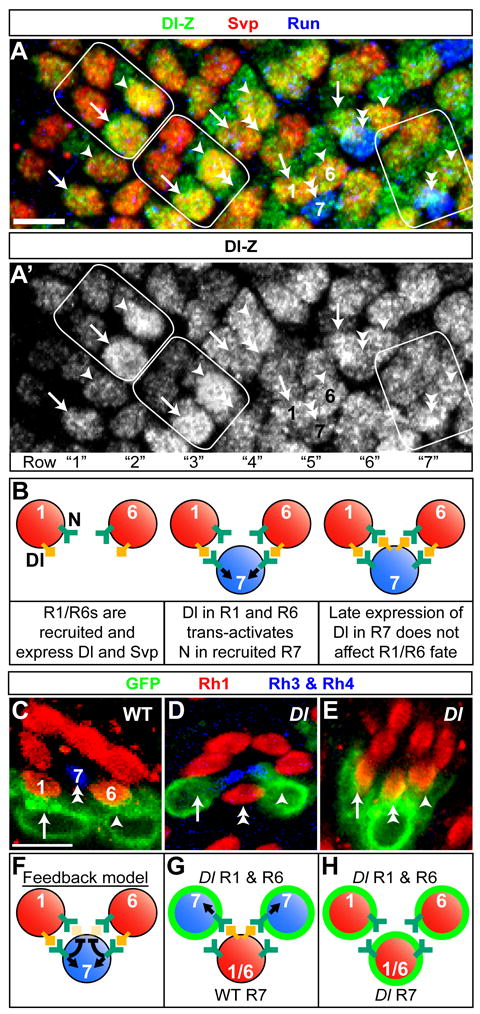
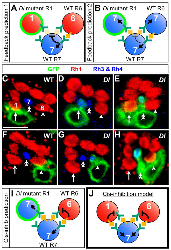
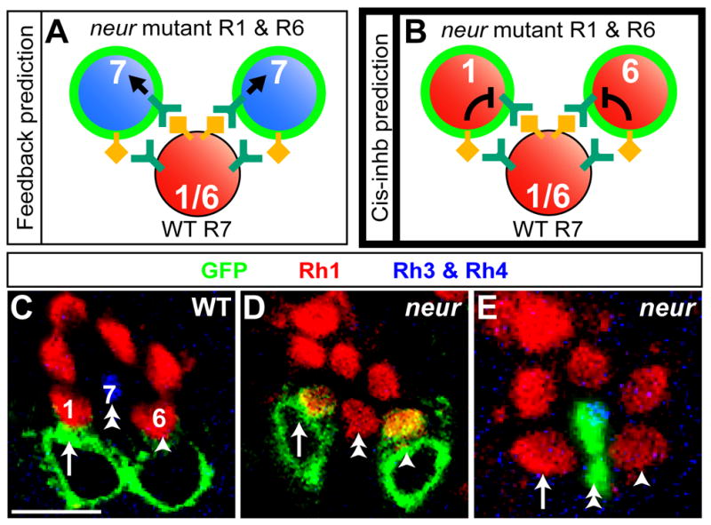
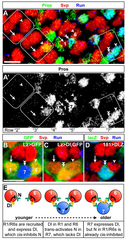
Lateral inhibition mediated by Delta/Notch (Dl/N) signaling is used throughout development to limit the number of initially equivalent cells that adopt a particular fate. Although adjacent cells express both Dl ligand and N receptor, signaling between them ultimately occurs in only one direction. Classically, this has been explained entirely by feedback: activated N can downregulate Dl, amplifying even slight asymmetries in the Dl or N activities of adjacent cells. Here, however, we present an example of lateral inhibition in which unidirectional signaling depends instead on Dl's ability to inhibit N within the same cell, a phenomenon known as cis-inhibition. By genetically manipulating individual R1/R6/R7 photoreceptor precursors in the Drosophila eye, we show that loss of Dl-mediated cis-inhibition reverses the direction of lateral signaling. Based on our finding that Dl in R1/R6s requires endocytosis to trans-activate but not to cis-inhibit N, we reexamine previously published data from other examples of lateral inhibition. We conclude that cis-inhibition generally influences the direction of Dl/N signaling and should therefore be included in standard models of lateral inhibition.


Figures

Figure 1
Figure 1. The R1 and R6 precursors normally express Dl first; if R1 and R6 lack Dl then the direction of signaling is reversed
(A,A′) A third instar larval (L3) eye disc heterozygous for a Dl-lacZ enhancer trap [32]. During late L3 a wave of differentiation passes from posterior (right) to anterior (left) across the eye disc, resulting in a gradient of ommatidial ages within a single disc: each vertical row of ommatidia is 1.5 hours older than the row to its immediate left. We arbitrarily define the row in which R1/R6 precursors first express Dl-lacZ as row “0” (Figure S1). R1 (arrow) and R6 (arrowhead) first express the the R1/R6- and R3/R4-specific transcription factor Seven-up (Svp; red) in row “1” (leftmost, unoutlined ommatidium) [25]. R7 precursors (double arrowheads) do not transcribe Dl (green) until row “3” (middle outlined ommatidium) (see Figure S1 for details). R7s ultimately express the R7- and R8-specific transcription factor Runt (Run; blue) [36]. Outlined ommatidia correspond to those in Figure 4A. Scale bar is 5 μm. (B) Schematic depicting the timing of Dl expression in wild-type R1/R6/R7 precursors. The three ommatidia depicted correspond to rows “1”, “2”, and “3”. (C–E) Mosaic adult ommatidia in which a small number of R1/R6/R7 precursors are homozygous for a particular chromosome arm (green; see Experimental Procedures). (C) an ommatidium in which both the R1 (arrow) and R6 (arrowhead) precursors are homozygous for a wild-type chromosome (FRT82): each has a large rhabdomere that remains in the outer part of the ommatidium and expresses the Rh1 rhodopsin (red) [37]. The wild-type R7 precursor (double arrowhead) has a small rhabdomere that extends into the center of the ommatidium and expresses Rh3 or Rh4 (blue) [37]. (D) When both the R1 (arrow) and R6 (arrowhead) precursors are homozygous Dl mutant then they have small central rhabdomeres that express Rh3 or Rh4 but not Rh1, indicating that they have adopted the R7 fate. The wild-type R7 precursor (double arrowhead) adopts the R1/R6 fate. (E) If Dl is also removed from the R7 precursor (double arrowhead) then Dl mutant R1 (arrow) and R6 (arrowhead) precursors no longer adopt the R7 fate and instead become R1/R6s. Scale bar is 5 μm. See Figure S2 for quantification. (F–H) Schematic representations of how the results in (C–E) can be explained by the classical feedback model of lateral inhibition. Precursor identities are indicated by position and black text. The fate each precursor adopts is indicated as in Figure 1B both by color (red = the R1/R6 fate and blue = R7 fate) and by the corresponding R cell fate number in white. Dl mutant cells are outlined in green, and Dl and N are depicted as in Figure 1B. (F) N in the R7 precursor is trans-activated by the early expression of Dl in the R1/R6 precursors. According to the feedback model, Dl in the R7 precursor is therefore downregulated, preventing it from later trans-activating N in the R1/R6 precursors. (G) When both R1/R6 precursors lack Dl, N is no longer activated in the R7 precursor. As a consequence, Dl in R7 is not downregulated and so can activate N in the R1/R6 precursors. The R1/R6 precursors therefore adopt the R7 fate, and the R7 precursor adopts the R1/R6 fate. (H) When Dl is removed from the R7 precursor, N is no longer activated in the Dl mutant R1/R6 precursors: all three precursors adopt the R1/R6 fate.
Figure 2
Figure 2. The phenotype caused by loss of Dl from the R1 or R6 precursor alone suggests that Dl cis-inhibits N in R1/R6 precursors
(A,B) The classical feedback model predicts two possible outcomes of removing Dl from the R1 precursor alone. Previous work has shown that the corresponding wild-type R6 precursor still activates N in the R7 precursor, which therefore adopts the R7 fate [14]. (A) The level of activated N in the R7 precursor may be sufficient to downregulate Dl. Both R1/R6 precursors will therefore adopt the R1/R6 fate. (B) Alternatively, a reduction in activated N within the R7 precursor may cause an increase in Dl that is sufficent to activate N in R1/R6 precursors. Both R1/R6 precursors will therefore adopt the R7 fate. (C–H) Mosaic adult ommatidia in which a small number of R1/R6/R7 precursors are homozygous for a particular chromosome arm (green) and are stained for their expression of Rh1 (red) and Rh3 or Rh4 (blue). Loss of Dl from the R1 (D; arrow) or R6 (G; arrowhead) precursor alone causes only that precursor to adopt the R7 fate (blue). This fate transformation depends on Dl from the R7 precursor (E and H, respectively). Scale bar is 5 μm. See Figure S2 for quantification. (I) Dl from the R7 precursor trans-activates N in the Dl mutant R1 precursor but does not trans-activate N in the wild-type R6 precursor. The only difference between the R1 and R6 precursors is that the latter expresses Dl. We therefore conclude that Dl inhibits N pathway activation. (J) Model: Dl in the R1/R6 precursors normally cis-inhibits N.
Figure 3
Figure 3. neur mutant R1/R6s do not trans-activate N in R7 and yet do not transduce the Dl signal from R7
(A,B) The outcomes predicted by the feedback and cis-inhibition models presented in Figures 1F and 2J, respectively. (A) The feedback model predicts that because Dl in neur mutant R1 and R6 precursors cannot trans-activate N in R7, Dl in R7 is not downregulated and will trans-activate N in the R1/R6 precursors, causing them to adopt the R7 fate. (B) The cis-inhibition model predicts that Dl in neur mutant R1/R6 precursors will still cis-inhibit N, preventing its trans-activation by Dl from the R7 precursor, and causing the neur mutant R1/R6 precursors to adopt the R1/R6 fate. (C–E) Mosaic adult ommatidia. Colors and scale bar are as in Figure 2. See Figure S2 for quantification. (C) A wild-type ommatidium in which R1 and R6 are homozygous wild-type (FRT82). (D) When both R1 (arrow) and R6 (arrowhead) are neur mutant, R7 (double arrowhead) adopts the R1/R6 fate, confirming that neur is required for Dl’s ability to trans-activate N. However, unlike Dl mutant R1/R6s, neur mutant R1/R6s never adopt the R7 fate, indicating that, as predicted by the cis-inhibition model, their N is not activated. (E) neur mutant R7s (double arrowhead) still adopt the R7 fate, confirming that loss of neur does not affect N’s ability to be trans-activated.
Figure 4
Figure 4. Cis-inhibition maintains the direction of signaling that is initially achieved by the ordered expression of Dl
(A,A′) An L3 eye disc in which approximately 11% of R1/R6/R7 precursors are homozygous Dl mutant. Because of Gal80 perdurance, homozygous clones are not yet marked (see Experimental Procedures). All R1 and R6 precursors initially express the R1/R6 marker Svp (red), but ultimately 14% (presumably those that are Dl mutant; see Figure S5 for details) instead express the R7-specific transcription factor Pros (green) as well as Run (blue). The outlined ommatidia correspond to those outlined in Figure 1A. In row “2” (leftmost outlined ommatidium), R1 (arrow) and R6 (arrowhead) express Svp only; at this time R7 does not yet express Dl-lacZ (see Figures 1A and S1). In row “3” (middle outlined ommatidium), a presumably Dl mutant R6 (arrowhead) expresses both the R1/R6 marker Svp and the R7 marker Pros; by this time the R7 has been recruited and first expresses Dl-lacZ (see Figures 1A and S1), although its nucleus is not yet apical enough to be visible in this plane. By row “7” (rightmost outlined ommatidium), the presumably Dl mutant R6 (arrowhead), like the R7 (double arrowhead) expresses Pros and Run but not Svp (as in Figure 1A, the R1 nucleus is below the plane of view). (B–D) L3 ommatidia labeled with antibodies against Svp (red) and Run (blue). (B) In lozenge-Gal4 (lz-Gal4), UAS-GFP heterozygotes, R1 and R6 express Svp and R7 expresses Run. Expression of GFP (green) initiates approximately simultaneously in progeny of the second mitotic wave (data not shown) [31]. (C) In lz-Gal4, UAS-Dl, UAS-GFP heterozygotes, many R7 precursors (double arrowhead) express Svp but not Run, indicating that they have adopted the R1/R6 fate. (D) In PM181-Gal4, UAS-Dl, UAS-lacZ heterozygotes, in which expression of beta-galactosidase (green) initiates in R7 precursors just after their recruitment (data not shown), R7 precursors never adopt the R1/R6 fate. In this ommatidium, PM181-Gal4 has been driving expression for approximately three hours; older R7s in which PM181-Gal4 has been expressed for more than twelve hours remain wild-type (data not shown). Scale bars are 5 μm. (E) Model. R1 and R6 precursors receive the EGF signal first and begin to differentiate and express Dl. The R7 precursor receives the EGF signal next and is immediately exposed to Dl in R1 and R6 before expressing any Dl of its own; its N is therefore trans-activated. By the time R7 expresses Dl, N in R1 and R6 has already been cis-inhibited by Dl and so cannot be trans-activated. We hypothesize that Dl in R7 cis-inhibits N but too late to prevent commitment to the R7 fate. However, in the interests of clarity, we have left this out of the schematic.

Comment in

Similar articles

Cited by

References

    1. Artavanis-Tsakonas S, Rand MD, Lake RJ. Notch signaling: cell fate control and signal integration in development. Science. 1999;284:770–776. - PubMed
    1. Baker NE. Notch signaling in the nervous system. Pieces still missing from the puzzle. Bioessays. 2000;22:264–273. - PubMed
    1. Bray SJ. Notch signalling: a simple pathway becomes complex. Nat Rev Mol Cell Biol. 2006;7:678–689. - PubMed
    1. Heitzler P, Simpson P. The choice of cell fate in the epidermis of Drosophila. Cell. 1991;64:1083–1092. - PubMed
    1. Wilkinson HA, Fitzgerald K, Greenwald I. Reciprocal changes in expression of the receptor lin-12 and its ligand lag-2 prior to commitment in a C. elegans cell fate decision. Cell. 1994;79:1187–1198. - PubMed

Publication types

MeSH terms

LinkOut - more resources