. 2009 Dec;98(4):356-66.
doi: 10.1016/j.ymgme.2009.06.016. Epub 2009 Jul 4.

Developmental expression pattern of the cholesterogenic enzyme NSDHL and negative selection of NSDHL-deficient cells in the heterozygous Bpa(1H)/+ mouse

Affiliations

Developmental expression pattern of the cholesterogenic enzyme NSDHL and negative selection of NSDHL-deficient cells in the heterozygous Bpa(1H)/+ mouse

David Cunningham et al. Mol Genet Metab. 2009 Dec.

Abstract

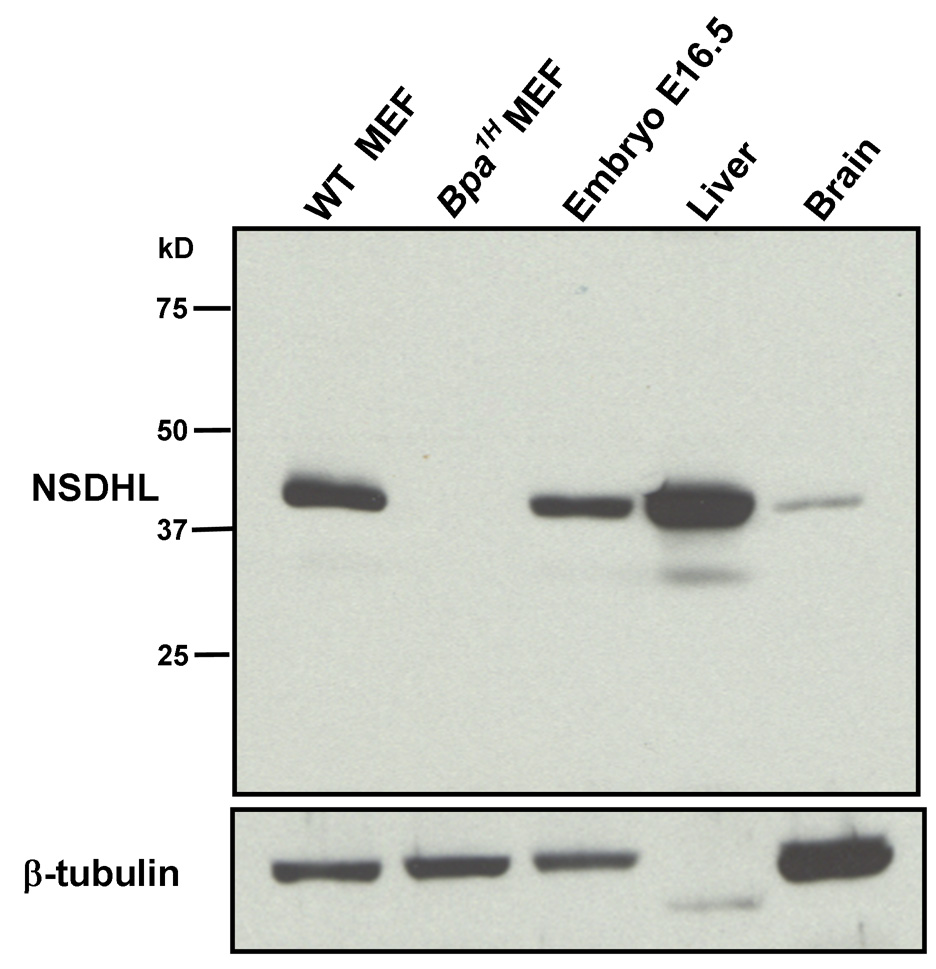
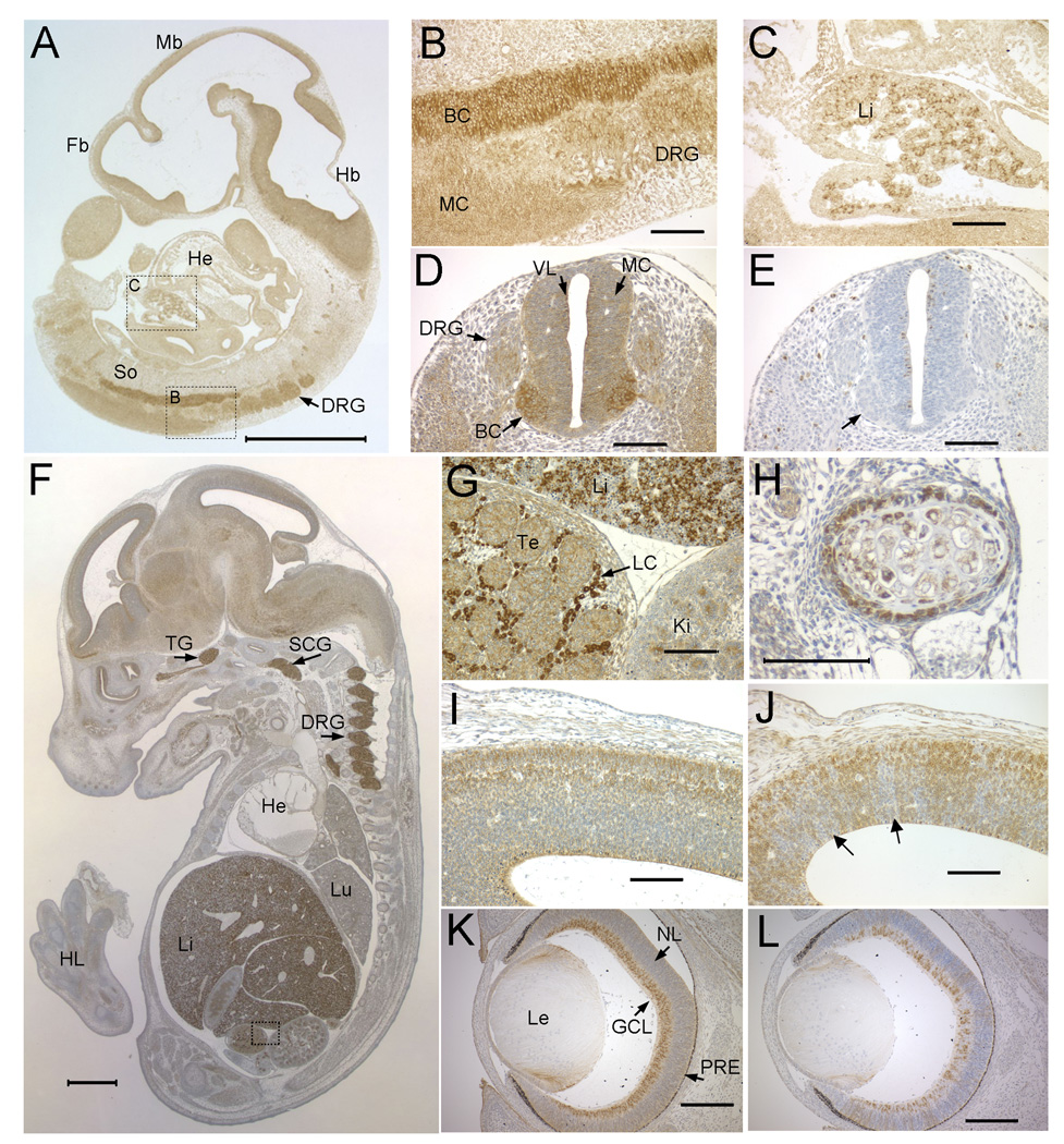
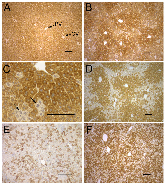
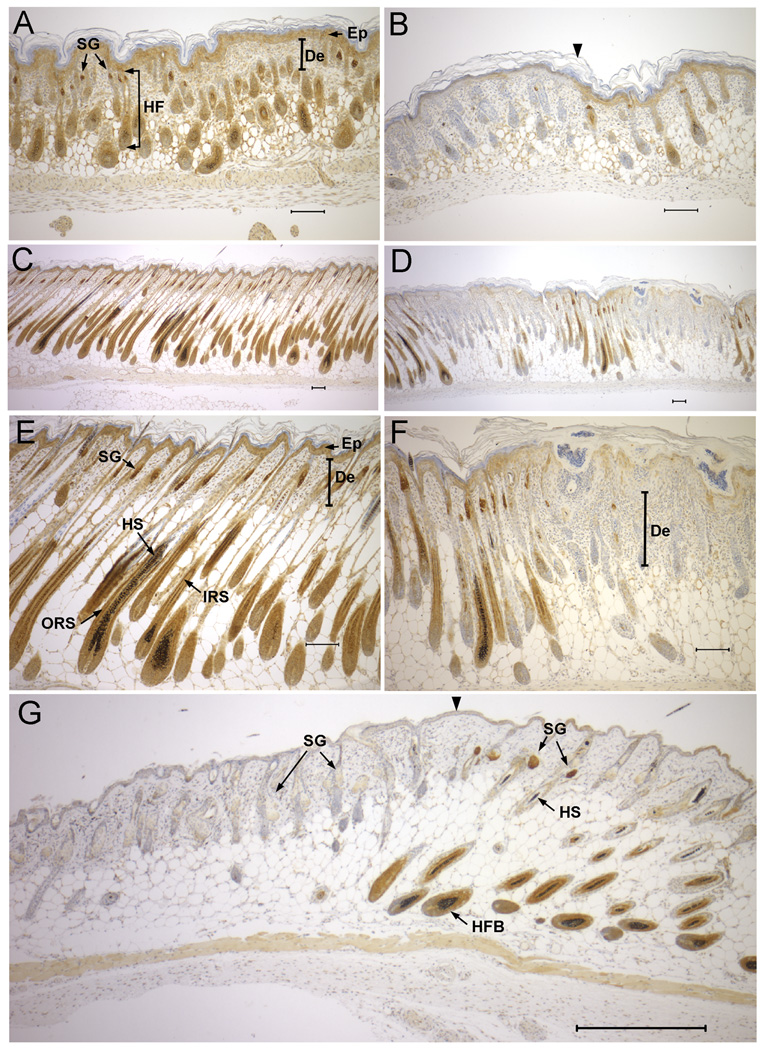
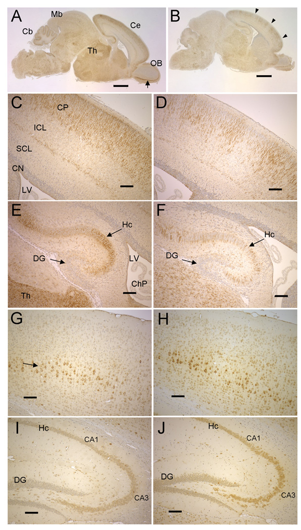
NSDHL (NAD(P)H sterol dehydrogenase-like), is a 3beta-hydroxysterol dehydrogenase thought to function in the demethylation of sterol precursors in one of the later steps of cholesterol biosynthesis. Mutations in the X-linked NSDHL gene cause CHILD syndrome in humans, and the male-lethal bare patches (Bpa) phenotype in mice. The relative level of NSDHL expression among different mouse tissues at several stages of embryogenesis and postnatal development was analyzed by immunohistochemistry. In wild type (WT) embryos, the highest levels of expression were seen in the liver, dorsal root ganglia, central nervous system, retina, adrenal gland and testis. Heterozygous Bpa(1H) females are mosaic for NSDHL expression due to normal random X-inactivation. NSDHL-deficient cells were detected in the developing cerebral cortex and retina of Bpa(1H) female embryos. In postnatal WT and Bpa(1H) animals, we compared the expression pattern of NSDHL in skin, an affected tissue; liver, a main site of cholesterol synthesis; and brain, a tissue dependent on endogenous synthesis of cholesterol due to lack of transport across the blood-brain barrier. Clonal populations of mutant cells were visible in the brain, skin and liver of Bpa(1H) pups. In the liver, the proportion of NSDHL negative cells dropped from approximately 50% at postnatal day 6 to approximately 20% at one year of age. In the brain, which showed the highest expression in cerebral cortical and hippocampal neurons, the proportion of NSDHL negative cells also dropped dramatically over the first year of life. Our results suggest that while NSDHL-deficient cells in the mosaic Bpa(1H) female are able to survive and differentiate during embryonic development, they are subject to negative selection over the life of the animal.


Figures

Figure 1
Figure 1. Detection of NSDHL in whole protein extracts by Western blot analysis using a purified anti-NSDHL polyclonal antibody
Approximately 10 µg of total protein from WT and Bpa1H MEFs, WT E16.5 embryo, 7 mo WT female liver and 7 mo WT female brain were resolved by PAGE, transferred to nylon membrane and probed with a polyclonal anti-NSDHL antibody at a 1:4000 dilution. Antibody binding was visualized by chemiluminescence detection (see methods). A single prominent band that migrated slightly above the 37 kD marker was detected in all samples except the Bpa1H MEF. A duplicate blot was probed with an anti-β-tubulin antibody to verify the presence of intact protein in the extracts.
Figure 2
Figure 2. Developmental expression pattern of NSDHL in mouse embryos
A. A saggital section from an E10.5 WT embryo immunostained for NSDHL showed the highest signal in the CNS and fetal liver. B. A higher magnification view of boxed area B in panel A showing intense staining of the basal column (BC) of the neural tube, with less signal in the mantle column (MC) and caudal dorsal root ganglia (DRG). C. A higher magnification view of boxed area C in panel A showing a subpopulation of strongly stained cells in the fetal liver (Li). D. A transverse section through the posterior neural tube illustrates the high level of NSDHL staining in the basal column of the neural tube relative to the mantle column and ventricular layer (VL). E. A section adjacent to that shown in panel D immunostained for phosphorylated histone H3, that marks mitotic cells, shows numerous dividing cells in the ventricular layer lining the neural tube, with no positive cells in the basal column, where the strongest NSDHL signal is seen (arrow). F. A saggital section from an E14.5 WT embryo immunostained for NSDHL shows intense staining in the liver, dorsal root ganglia, and trigeminal ganglion (TG). Signal in the brain is higher than in other tissues such as heart (He) and lung (Lu). G. A high magnification view of the boxed area from panel F, showing strong staining of the Leydig cells (LC) in the testis, comparable to the level in hepatocytes seen in the adjacent fetal liver. H. High magnification view of developing rib from section shown in panel F. NSDL staining is seen in cells of the condensing mesenchyme surrounding the chondrocytes of the rib. I. A high magnification view of the posterior neopallial cortex of the brain from the section shown in panel F. J. The same region of the brain shown in panel I from a Bpa1H E14.5 female embryo was immunostained for NSDHL. Note the patchy staining pattern, with radiating sectors of neurons showing no staining (arrows). K. A transverse section through the eye of a WT E15.5 embryo showing strong NSDHL staining in the ganglion cell layer (GCL). L. NSDHL staining of a Bpa1H E15.5 eye, showing mosaic expression in the GCL with radiating sectors of NSDHL positive and negative cells in progeny cells of the developing retina. Sections in panels B–L were lightly counterstained with hematoxylin. The size bars in panels A and F represent 1 mm. Size bars in all other panels denote 100 µm. Abbreviations: BC, basal column; DRG, dorsal root ganglion; Fb, forebrain; GCL, ganglion cell layer; Hb, hindbrain; He, heart; HL, hind limb; Ki, kidney; LC, Leydig cell; Le, lens; Lu, lung; Mb, midbrain; MC, mantle column; NL, neuroblast layer; PRE, pigmented retinal epithelium; VL, ventricular layer.
Figure 3
Figure 3. Immunostaining of NSDHL in WT and Bpa1H liver
A. Adult (3 mo) WT liver shows strong staining thoughout the lobule, with slightly higher signal in the pericentral region. B. An adult (13 mo) Bpa1H liver with a large majority of NSDHL positive cells. C. A high magnification view of the liver shown in panel B showing small clusters of hepatocytes that did not stain for NSDHL. D. An adult (13 mo) Bpa1H liver with a relatively large number of NSDHL negative hepatocytes. Note the high proportion of NSDHL positive cells surrounding the central veins. NSDHL staining in the liver of a P6 Bpa1H female. F. NSDHL staining in the liver of a P25 Bpa1H female. Abbreviations: CV, central vein; PV, portal vein. Size bars represent 100 µm.
Figure 4
Figure 4. NSDHL expression in developing dorsal skin and hair follicles
A. NSDHL staining in dorsal skin of a P2 WT pup shows strong signal in the sebaceous glands, and expression in the inner root sheath of the developing hair follicle, as well as the epidermis. B. In dorsal skin of a Bpa1H P2 pup, regions lacking detectable NSDHL in the epidermis and developing hair follicles (left of arrowhead) appeared similar histologically to regions that stained positively for NSDHL (right of arrowhead). C. WT skin from P6 displayed strong NSDHL staining in the outer root sheath and moderate staining in the inner root sheath of the maturing hair follicle. D. In Bpa1H skin at P6, NSDHL negative regions showed a thickening of the epidermis and dermis with developmentally delayed hair follicles. E. Higher magnification view of the sample shown in panel C. F. Higher magnification of the sample shown in panel D. G. NSDHL staining in dorsal skin of a Bpa1H female at 8 weeks of age. Strong staining in sebaceous glands and the inner root sheath of anagen stage hair follicles is visible in the region to the right of the arrowhead. The area on the left shows virtually no staining in the enlarged sebaceous glands and mainly telogen stage hair follicles. No hair shafts are present within the hair follicles in this region. Abbreviations: De, dermis; Ep, epidermis; HF, hair follicle; HFB, hair follicle bulb; HS, hair shaft; IRS, inner root sheath; ORS, outer root sheath; SG, sebaceous gland. Size bars represent 100 µm.
Figure 5
Figure 5. NSDHL in early postnatal and adult brains of WT and Bpa1H females
A. NSDHL staining of a saggital section from a WT P2 brain. The arrow indicates the mitral layer of the olfactory bulb. B. NSDHL staining of a Bpa1H P2 brain. Arrowheads indicate radial sectors of low NSDHL signal in the cerebrum. C. A higher magnification of the cerebral cortex from the section shown in panel A shows a broad, even band of positively stained pyramidal neurons in the outer layer of the cortex. D. A higher magnification of the cerebral cortex from the Bpa1H brain section shown in panel B reveals regions of unstained cortical neurons that were not seen in the WT brain. E. A higher magnification of the hippocampus (Hc) from the WT brain shown in panel A. F. High magnification of the hippocampus from the Bpa1H brain shown in panel B, showing a mosaic pattern of NSDHL within the hippocampus. G. NSDHL staining in the cerebral cortex on a saggital section of a WT adult brain. The arrow indicates a layer of relatively large pyramidal neurons that showed the highest level of NSDHL signal in the adult cerebral cortex. H. NSDHL staining in the cerebral cortex of a Bpa1H adult brain. I. NSDHL staining in the hippocampus of a WT adult brain. J. NSDHL staining in the hippocampus of a Bpa1H adult brain. Abbreviations: Cb, cerebellum; Ce, cerebrum; ChP, choroid plexus; CN, cortical neurepithelium; CP, cortical plate; Cx, cerebral cortex; DG, dentate gyrus; Hc, hippocampus; Ht, hypothalamus; ICL, intermediate cortical layer; LV, lateral ventricle; OB, olfactory bulb; SCL, subventricular cortical layer; Th, thalamus. Size bars in panels A and B correspond to 1 mm. Size bars in panels C–J correspond to 100 µm.

References

    1. Simons K, Toomre D. Lipid rafts and signal transduction. Nat Rev Mol Cell Biol. 2000;1:31–39. - PubMed
    1. Ikonen E. Cellular cholesterol trafficking and compartmentalization. Nat Rev Mol Cell Biol. 2008 Sep;:125–138. - PubMed
    1. Jeong J, McMahon AP. Cholesterol modification of Hedgehog family proteins. J Clin Invest. 2002;110:591–596. - PMC - PubMed
    1. Wassif CA, Zhu P, Kratz L, Krakowiak PA, Battaile KP, Weight FF, Grinberg A, Steiner RD, Nwokoro NA, Kelley RI, Stewart RR, Porter FD. Biochemical, phenotypic and neurophysiological characterization of a genetic mouse model of RSH/Smith--Lemli--Opitz syndrome. Hum Mol Genet. 2001;10:555–564. - PubMed
    1. Ohashi K, Osuga J, Tozawa R, Kitamine T, Yagyu H, Sekiya M, Tomita S, Okazaki H, Tamura Y, Yahagi N, Iizuka Y, Harada K, Gotoda T, Shimano H, Yamada N, Ishibashi S. Early embryonic lethality caused by targeted disruption of the 3-hydroxy-3-methylglutaryl-CoA reductase gene. J Biol Chem. 2003;278:42936–42941. - PubMed

Publication types

MeSH terms