

Desmoplastic ameloblastoma - A review
- PMID: 19631576
- PMCID: PMC6022750
- DOI: 10.1016/j.oraloncology.2009.01.016
Desmoplastic ameloblastoma - A review
Abstract
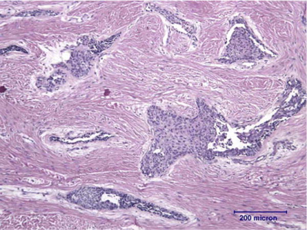
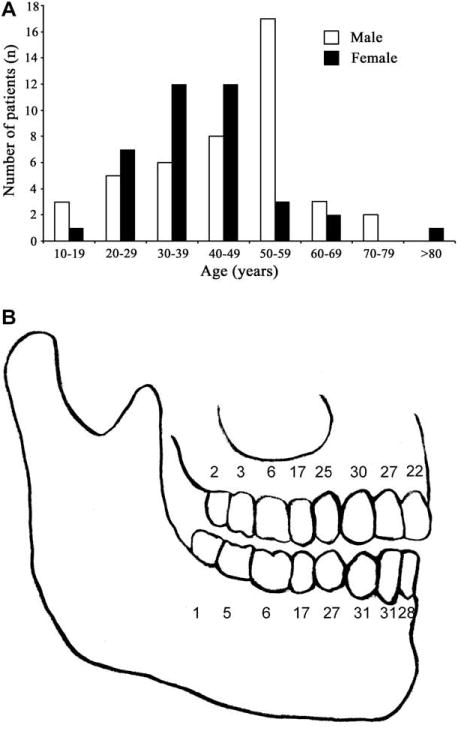
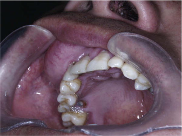
Among the ameloblastomas, the desmoplastic variation is rare. The desmoplastic ameloblastoma (DA) is characterized by specific clinical, imaging, and histological features. The here presented retrospective analysis investigated the clinicoradiographic features of an overall of 115 DA-cases, having been reported in literature from 1984 to 2008. DA showed a nearly equal male to female ratio (55/59) with a prevalence within the forth and fifth decades. Sixty-two lesions occurred in the mandible and fifty-one lesions in the maxilla. Clinically, a painless swelling with buccal extension was the most common presentation being found in 48 cases. Radiologically, the lesion often presented multilocular (49.3%; 36/73), mixed radiolucent/radiopaque (55.6%; 50/90) and with ill-defined borders (64.0%; 48/75). Whereas enucleation provided a recurrence rate of 21.1%, resection reduced this rate remarkably to 3.1%. The average period until recurrence was 36.9 months. Histologically, scattered epithelial nests and extensively desmoplasia were prominent features of DA. In conclusion, these retrospective results confirm the statement that DA is a variation among ameloblastomas. DA present clinicoradiographic and histologic distinct features, when compared with "conventional ameloblastomas".
Conflict of interest statement
Conflict of Interest Statement
None declared
Figures

References
-
- Waldron CA, el-Mofty SK. A histopathologic study of 116 ameloblastomas with special reference to the desmoplastic variant. Oral Surg Oral Med Oral Pathol. 1987;63(4):441–51. - PubMed
-
- Smullin SE, Faquin W, Susarla SM, Kaban LB. Peripheral desmoplastic ameloblastoma: report of a case and literature review. Oral Surg Oral Med Oral Pathol Oral Radiol Endod. 2008;105(1):37–40. - PubMed
-
- Philipsen HP, Reichart PA, Takata T. Desmoplastic ameloblastoma (including “hybrid” lesion of ameloblastoma). Biological profile based on 100 cases from the literature and own files. Oral Oncol. 2001;37(5):455–60. - PubMed
-
- Eversole LR, Leider AS, Strub D. Radiographic characteristics of cystogenic ameloblastoma. Oral Surg Oral Med Oral Pathol. 1984;57(5):572–7. - PubMed
-
- Gardner DG, Heikinheimo K, Shear M, Philipsen HP, Coleman H. Ameloblastomas. In: Barnes L, Eveson EJ, Reichart P, Sidransky D, editors. World Health Organization classification of tumors: pathology and genetics of head and neck tumors. 3rd. Lyon: IARC Press; 2005. pp. 296–300.
Publication types
MeSH terms
Grants and funding
LinkOut - more resources
Full Text Sources
