. 2009 Oct 1;334(1):119-32.
doi: 10.1016/j.ydbio.2009.07.023. Epub 2009 Jul 23.

VEGF-A and Semaphorin3A: modulators of vascular sympathetic innervation

Affiliations

VEGF-A and Semaphorin3A: modulators of vascular sympathetic innervation

Jennifer B Long et al. Dev Biol. .

Abstract

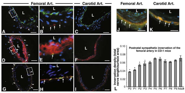
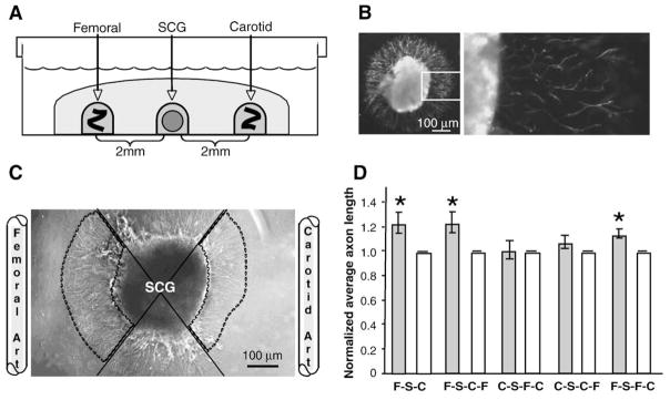
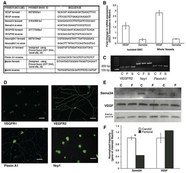
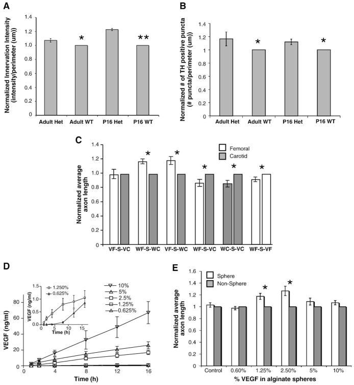
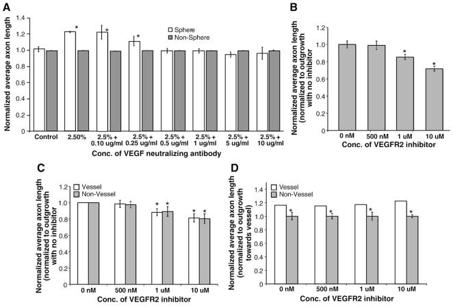
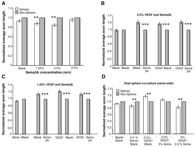
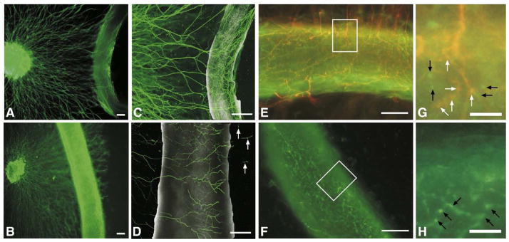
Sympathetic nerve activity regulates blood pressure by altering peripheral vascular resistance. Variations in vascular sympathetic innervation suggest that vascular-derived cues promote selective innervation of particular vessels during development. As axons extend towards peripheral targets, they migrate along arterial networks following gradients of guidance cues. Collective ratios of these gradients may determine whether axons grow towards and innervate vessels or continue past non-innervated vessels towards peripheral targets. Utilizing directed neurite outgrowth in a three-dimensional (3D) co-culture, we observed increased axon growth from superior cervical ganglion explants (SCG) towards innervated compared to non-innervated vessels, mediated in part by vascular endothelial growth factor (VEGF-A) and Semaphorin3A (Sema3A) which both signal via neuropilin-1 (Nrp1). Exogenous VEGF-A, delivered by high-expressing VEGF-A-LacZ vessels or by rhVEGF-A/alginate spheres, increased sympathetic neurite outgrowth while exogenous rhSema3A/Fc decreased neurite outgrowth. VEGF-A expression is similar between the innervated and non-innervated vessels examined. Sema3A expression is higher in non-innervated vessels. Spatial gradients of Sema3A and VEGF-A may promote differential Nrp1 binding. Vessels expressing high levels of Sema3A favor Nrp1-PlexinA1 signaling, producing chemorepulsive cues limiting sympathetic neurite outgrowth and vascular innervation; while low Sema3A expressing vessels favor Nrp1-VEGFR2 signaling providing chemoattractive cues for sympathetic neurite outgrowth and vascular innervation.


Figures

Fig. 1
Fig. 1
Localization of murine vascular sympathetic innervation. (A–C) Immunolabeling for PECAM-1 (green) to mark vascular endothelial cells, tyrosine hydroxylase (red) to mark sympathetic nerves and DAPI (blue) to mark nuclei in femoral (A, B=boxed area in panel A) and carotid arteries (C). Localization of both tyrosine hydroxylase and PECAM-1 is found in adult femoral artery sections (A and B, arrows) but tyrosine hydroxylase localization is absent from adult carotid artery sections (C). L=lumen. Scale bar=50 μm. (D–F) Immunolabeling for smooth muscle cell actin (red) to mark vascular SMCs, tyrosine hydroxylase (green) and DAPI (blue) to mark nuclei in femoral (D, E=boxed area in panel D) and carotid arteries (F). Innervation patterns match that of PECAM-1/TH double-labeling (D and E, arrows). Sympathetic nerves are localized to the medio-adventitial border of the femoral arteries (orange–yellow fluorescence — arrows). Scale bar=50 μm. (G–I) Immunolabeling for synaptophysin (red) to mark synapses, tyrosine hydroxylase (green) to mark sympathetic nerves and DAPI (blue) to mark nuclei in femoral (G, H=boxed area in panel G) and carotid (I) artery sections. Both vessels show significant synapse formation around the periphery, but synapses with sympathetic nerves are localized only in femoral artery sections (G and H, orange–yellow fluorescence — arrows). Scale bar=50 μm. (J and K) Immunolabeling for synaptophysin (red) to mark synapses, vesicular acetylcholine transferase (green) to mark sympathetic nerves and DAPI (blue) to mark nuclei in femoral (J) and carotid (K) artery sections. Both vessels show significant synapse formation around the periphery (orange–yellow fluorescence — arrows). Scale bar=50 μm. (L) Innervation densities were quantified in femoral arteries from postnatal days 0 through 9 mice by whole mount tyrosine hydroxylase immunofluorescence. Innervation density was calculated as described in Materials and methods and represents total nerve length (μm)/surface area (μm2) for the superficial half of each vessel. Significant differences between postnatal and adult innervation differences exist between days 0–3, after which no differences exist. Carotid arteries are not innervated (data not shown) (n=6; vertical bars represent standard error; *=p<0.05).
Fig. 2
Fig. 2
Directed migration in neurovascular co-cultures. (A) Schematic representing three-dimensional in-vitro co-culture. Whole SCG explants from postnatal day 2 mice were co-cultured in the presence of adult femoral and carotid artery segments. Tissues were embedded in type I collagen gel 2 mm apart and cultured for 16 h. (B) Tyrosine hydroxylase immunolabeling in whole SCG explants. Left, low magnification; right (boxed inset), high magnification. Scale bar=100 μm. (C) Representative image of SCG co-cultured with femoral and carotid artery segments. Femoral and carotid artery segments were placed 2 mm from SCG explants. To quantify outgrowth, the area occupied by the SCG and its neurites was divided into quadrants (solid lines). Average axon length was measured for 25 axons in the femoral and carotid directed quadrants (outlined by dashed lines). Carotid directed average axon length was normalized to 1 and femoral/carotid ratios were calculated. Scale bar=100 μm. (D) Directed neurite outgrowth of sympathetic axons from whole SCG explants. Neurites of equal average length grew radially from SCG alone controls (data not shown). In femoral/carotid co-cultures (first pair of bars) (FSC) the average axon length was significantly increased towards the femoral compared to the carotid artery. (n=14; *=p<0.05; vertical bars represent standard error.) To test the effect of vessel rearrangements on directed neurite outgrowth, SCG explants (S) were cultured in the presence of various arrangements of vessels. When femoral segments (F) were placed on the same side as carotid segments (C), either proximal (second pair of bars) (FSCF) or distal to the SCG (S) (third pair of bars) (CSFC) outgrowth towards the femoral segment was blunted, with the placement of carotid segments disrupting the typical increased outgrowth observed towards femoral segments. (n=14; & =p<0.05; vertical bars represent standard error.) When femoral segments (F) were placed on the same side as carotid segments (C), either distal (fourth pair of bars) (CSCF) or proximal to the SCG (S) (fifth pair of bars) (FSFC) outgrowth towards the carotid segment was unchanged.
Fig. 3
Fig. 3
Expression of guidance cues and their receptors in vessels and SCG. (A) VEGF-A, Sema3A, VEGFR2, Nrp1 and PlexinA1 forward and reverse primer sets used for quantitative real-time PCR and RT-PCR. (B) Quantitative real-time PCR analysis of VEGF-A and Sema3A in femoral and carotid artery isolated smooth muscle cells (SMC) and whole vessels. Femoral arteries and SMC express a three-fold increase in VEGF-A mRNA compared to carotid arteries and SMC while the expression profile of Sema3A in the reciprocal. (n=3 for each gene of interest; *=p<0.05; vertical bars represent standard error.) (C) RT-PCR analysis of receptor expression in vessels and SCG. Femoral and carotid arteries as well as SCG express mRNA for VEGFR2, Nrp1 and PlexinA1. (D) Immunofluorescent analysis of VEGFR1, VEGFR2, PlexinA1 and Nrp1 in postnatal day 2 dissociated SCG neurons. Scale bar=50 μm. (E) Protein expression of guidance cues in postnatal day 2 whole vessel lysates. Representative Western blots of femoral (F) and carotid (C) artery segments identifying Sema3A and VEGF-A normalized to beta actin. (F) Quantification of Western blot data. Sema3A expression is significantly higher in carotid compared to femoral artery segments while VEGF-A expression is not significantly different between vessels (n=3 for each gene of interest; *=p<0.05; vertical bars represent standard error).
Fig. 4
Fig. 4
The effect of VEGF-A on directed neurite outgrowth (unless otherwise noted n=14, vertical bars represent standard error). (A, B) Innervation analysis of sympathetic innervation in VEGF-A (hi/+) compared to wild-type littermates. Tyrosine hydroxylase immunostaining revealed significant increases in sympathetic innervation density measured by both immunofluorescence intensity (A) and # of TH positive puncta (B) in both adult and postnatal day 16 compared to matched WT controls. (n=5 for each age group; *= p<0.05, **= p<0.01.) (C) Neurovascular co-cultures using vessels from VEGF-A (hi/+) heterozygous mice and wild-type littermates. When placed on either side of SCG, VEGF-A (hi/+) carotid and femoral artery segments elicited an increased equal neurite outgrowth compared to wild-type carotid segments, reflecting increased VEGF-A expression in both vessel segments (first pair of bars), in contrast to the differential neurite outgrowth towards femoral artery segments observed when wild-type vessel segments were used (second pair of bars). Similar outgrowth was noted when VEGF-A (hi/+) femoral artery segments (VF) were compared to wild-type carotid artery segments (WC) (third pair of bars). VEGF-A (hi/+) carotid artery segments (VC) promoted increased average axon length from the SCG (S) when cultured with both wild-type femoral (WF) (fourth pair of bars) and wild-type carotid (WC) (fifth pair of bars) vessel segments. Lastly, VEGF-A (hi/+) (VF) femoral segments elicited increased outgrowth compared to wild-type femoral artery segments (WF) (sixth pair of bars). Interestingly, femoral artery segments from VEGF-A (hi/+) (VF) mice did not promote further increases in expected average axon length when cultured with wild-type carotid artery segments (WC) than did wild-type femoral artery segments (WF) (compare the second and third pairs of bars). (*=p<0.05.) (D) Release of VEGF-A into type I collagen gel from VEGF-A/alginate spheres by ELISA. VEGF-A/alginate spheres released increasing amounts of VEGF-A into the media over 16 h. Amounts of VEGF-A released ranged from 70 ng/ml (10% w/v VEGF-A) to 1.5 ng/ml (1.25% and 0.625% w/v VEGF-A, inset). (E) SCG outgrowth in response to VEGF-A/alginate spheres. Average sympathetic neurite lengths towards 2.5% and 1.25% w/v VEGF-A/alginate spheres was approximately 25% and 18% higher respectively compared to the non-vessel quadrant. Control, 0.625%, 5% and 10% w/v spheres caused no significant increases in outgrowth. (*=p<0.05.)
Fig. 5
Fig. 5
The effect of VEGF-A neutralization on directed neurite outgrowth (n=14, vertical bars represent standard error). (A) The effect of VEGF-A neutralizing antibodies on average axon length. Anti-human VEGF-A antibodies were used to neutralize the effect of VEGF-A released from alginate spheres. Concentrations as low as 500 μg/ml were able to block the effect of the VEGF-A/alginate spheres compared to sphere alone controls. (*=p<0.05.) (B) The effect of a VEGFR2 inhibitor on average axon length. A highly specific cell permeable VEGFR2 tyrosine kinase inhibitor was used to block signaling from VEGF-A released from alginate spheres. Concentrations as low as 1 μg/ml were able to block the effect of VEGF-A/alginate spheres compared to sphere alone controls (first bar on the left). (C) The effect of the VEGFR2 inhibitor on femoral–SCG neurovascular co-cultures. The cell permeable VEGFR2 inhibitor was also able to decrease neurite outgrowth elicited by femoral artery segments co-cultured with SCG. (*=p<0.05.) Average axon length (for vessel and non-vessel cultures) at all inhibitor concentrations is normalized to axon lengths with no VEGFR2 inhibitors. On both the vessel and non-vessel sides of the SCG, there is a decreased average axon length compared to the no inhibitor control (first pair of bars on the left). (D) The effect of VEGFR2 inhibitors on the increase in axon outgrowth elicited by femoral artery segments. At each concentration of VEGFR2 inhibitor, average axon length on the non-vessel side of the SCG is normalized to the average axon length towards the vessel. Although VEGFR2 tyrosine kinase inhibitors were able to significantly decrease overall axon length in femoral–SCG neurovascular co-cultures at all inhibitor concentrations tested, the ratio of vessel/non-vessel outgrowth was maintained with significantly more outgrowth observed in the quadrant towards the femoral artery segment compared to the non-vessel quadrant. (*=p<0.05.)
Fig. 6
Fig. 6
The effect of Sema3A on directed neurite outgrowth (n=14, vertical bars represent standard error). (A) SCG outgrowth in response to Sema3A/alginate spheres. An average sympathetic neurite length towards 1.25% and 2.5% Sema3A/alginate spheres was decreased by approximately 25% compared to empty spheres. 5% Sema3A/alginate spheres caused no significant decrease in neurite outgrowth. (**=p<0.01.) (B, C) SCG outgrowth in response to VEGF-A/alginate and Sema3A/alginate spheres placed on opposite sites of the SCG. Average neurite outgrowth towards VEGF-A spheres was 17% higher compared to empty spheres and was decreased by 21% towards Sema3A spheres compared to empty spheres. There were no significant differences when VEGF-A and Sema3A spheres were used in combination in dual sphere cultures compared to single sphere cultures at either 2.5% (B) or 1.25% (C) VEGF and Sema3A. (**=p<0.001.) (D) SCG outgrowth in response to VEGF-A/Sema3A/Blank alginate spheres. Alginate spheres were placed in combination on the same side of the SCG. Neurite outgrowth towards VEGF-A/Blank spheres was 16.5% higher and towards the Sem3A/Blank spheres was 14% lower compared to the non-sphere side. When 2.5% VEGF-A and a 5% Sema3A spheres were used, there was only a 6% increase in outgrowth towards the spheres compared to the non-sphere side. When 5% VEGF-A and 2.5% Sema3A spheres were used, there was an 18% decrease in outgrowth compared to the non-sphere side. (*=p<0.05.)
Fig. 7
Fig. 7
Five-day neurovascular co-culture. (A, B) Fluorescent images of tyrosine hydroxylase sympathetic axons growing towards femoral (A) or carotid (B) artery segments for five days in co-culture. In femoral–SCG co-cultures axons grew out to, but did not pass the vessel in 6/7 co-cultures. In addition, the neurons appeared to make contact with and wrap around the vessel, forming a network parallel and perpendicular to the axis of the vessel similar to that seen in ex-vivo whole mount preparations (data not shown). Scale bar=200 μm. (C, D) Confocal images of tyrosine hydroxylase immunofluorescence of SCG explants cultured with femoral (C) or carotid (D) artery segments for five days in 2.5 mg/ml collagen. Neurons comprising the femoral network were arranged both parallel and perpendicular to the vessel axis, investing the vessel, while neurons comprising the carotid network were arranged mainly perpendicular to the vessel axis with occasional axons migrating past the vessel segment (arrows) in 5/7 co-cultures. Scale bar=200 μm. (E, F) Merged images of a five-day co-culture of a femoral artery segment with a SCG (E) and a segment of femoral artery cultured alone (F), illustrating synaptophysin staining (green fluorescence) and tyrosine hydroxylase staining (red fluorescence) of presynaptic varicosities, revealing co-localization of a portion of the varicosities suggestive of sympathetic re-innervation (orange fluorescence) in (E) and no detectable tyrosine hydroxylase staining in the mono-cultured femoral artery segment (F). Scale bar=100 μm. (G, H) High power magnification insets of (E) and (F) illustrating specific synaptophysin labeled varicosities (green fluorescence denoted by black arrows) and synaptophysin/tyrosine hydroxylase co-labeled varicosities (orange fluorescence denoted by white arrows) in (G) and specific synaptophysin labeled varicosities (green fluorescence denoted by black arrows) in the monoculture in(H). Scale bar=25 μm.
Fig. 8
Fig. 8
Working model for the role of vascular-derived VEGF-A and Sema3A in modulating sympathetic axon outgrowth. (A) Schematic representing our model for the roles of VEGF-A and Sema3A in modulating sympathetic outgrowth. Both VEGF-A and Sema3A bind to Nrp1 and promote its association with a co-receptor, either VEGFR2 or PlexinA1. Both femoral and carotid arteries produce VEGF-A. (B) The femoral artery was noted to produce slightly more VEGF-A than the carotid artery. In the absence of chemorepulsive signals, femoral-derived VEGF-A can bind to Nrp1, which can then associate with VEGFR2, resulting in increased neurite outgrowth. (C) In contrast, the carotid artery was noted to produce approximately three times more Sema3A than the carotid artery, favoring Nrp1-bound Sema3A to associate with PlexinA1, resulting in blunted neurite outgrowth.

Similar articles

Cited by

References

    1. Adams RH, Wilkinson GA, Weiss C, Diella F, Gale NW, Deutsch U, Risau W, Klein R. Roles of ephrinB ligands and EphB receptors in cardiovascular development: demarcation of arterial/venous domains, vascular morphogenesis, and sprouting angiogenesis. Genes Dev. 1999;13:295–306. - PMC - PubMed
    1. Appleton BA, Wu P, Maloney J, Yin J, Liang WC, Stawicki S, Mortara K, Bowman KK, Elliott JM, Desmarais W, Bazan JF, Bagri A, Tessier-Lavigne M, Koch AW, Wu Y, Watts RJ, Wiesmann C. Structural studies of neuropilin/antibody complexes provide insights into semaphorin and VEGF binding. EMBO J. 2007;26:4902–4912. - PMC - PubMed
    1. Bagnard D, Vaillant C, Khuth ST, Dufay N, Lohrum M, Puschel AW, Belin MF, Bolz J, Thomasset N. Semaphorin 3A-vascular endothelial growth factor-165 balance mediates migration and apoptosis of neural progenitor cells by the recruitment of shared receptor. J Neurosci. 2001;21:3332–3341. - PMC - PubMed
    1. Belliveau DJ, Krivko I, Kohn J, Lachance C, Pozniak C, Rusakov D, Kaplan D, Miller FD. NGF and neurotrophin-3 both activate TrkA on sympathetic neurons but differentially regulate survival and neuritogenesis. J Cell Biol. 1997;136:375–388. - PMC - PubMed
    1. Birch DJ, Turmaine M, Boulos PB, Burnstock G. Sympathetic innervation of human mesenteric artery and vein. J Vasc Res. 2008;45:323–332. - PubMed

Publication types

Substances