. 2009 Nov 27;158(1-3):26-31.
doi: 10.1016/j.regpep.2009.07.010. Epub 2009 Jul 23.

Bombesin enhances TGF-beta growth inhibitory effect through apoptosis induction in intestinal epithelial cells

Affiliations

Bombesin enhances TGF-beta growth inhibitory effect through apoptosis induction in intestinal epithelial cells

Xianghua Liu et al. Regul Pept. .

Abstract

Mammalian intestinal epithelium undergoes continuous cell turn over, with cell proliferation in the crypts and apoptosis in the villus. Both transforming growth factor (TGF)-beta and gastrin-releasing peptide (GRP) are involved in the regulation of intestinal epithelial cells for division, differentiation, adhesion, migration and death. Previously, we have shown that TGF-beta and bombesin (BBS) synergistically induce cyclooxygenase-2 (COX-2) expression and subsequent prostaglandin E(2) (PGE2) production through p38(MAPK) in rat intestinal epithelial cell line stably transfected with GRP receptor (RIE/GRPR), suggesting the interaction between TGF-beta signaling pathway and GRPR. The current study examined the biological responses of RIE/GRPR cells to TGF-beta and BBS. Treatment with TGF-beta1 (40 pM) and BBS (100 nM) together synergistically inhibited RIE/GRPR growth and induced apoptosis. Pretreatment with SB203580 (10 microM), a specific inhibitor of p38(MAPK), partially blocked the synergistic effect of TGF-beta and BBS on apoptosis. In conclusion, BBS enhanced TGF-beta growth inhibitory effect through apoptosis induction, which is at least partially mediated by p38(MAPK).


Figures

Figure 1
Figure 1. TGF-β and BBS inhibited cell growth in RIE/GRPR cells
The cells were seeded in 12-well plates for 24 h, then treated with TGF-β1 (40 pM), BBS (100 nM), or TGF-β1 (40 pM) and BBS (100 nM) together. Cell numbers were obtained from triplicate wells and expressed as mean±SEM. *p < 0.05 compared with the group of vehicle control. † p<0.05 compared with either TGF-β1 or BBS alone.
Figure 2
Figure 2. TGF-β and BBS synergistically induced apoptosis in RIE/GRPR cells
A. The cells were seeded in 12-well plates for 24 h, then treated with TGF-β1 (40 pM), BBS (100 nM), or TGF-β1 (40 pM) and BBS (100 nM) together for 24 h. A. DNA fragmentation was quantified by a cell death detection assay. B. Cells were harvested and stained with Annexin V-FITC and propidium iodide. Apoptotic cells with Annexin V-FITC only were expressed as mean±SEM. *p < 0.05 compared with the group of vehicle control, † p < 0.05 compared with the group with treatment of TGF- β alone.
Figure 3
Figure 3. TGF-β and BBS induced cell cycle arrest in RIE/GRPR cells
The cells were seeded for 24 h, then starved for 48 h. The cells were treated with TGF-β1 (40 pM), BBS (100 nM), or TGF-β1 (40 pM) and BBS (100 nM) together for 24 h. The cells were pulsed with BrdU during the last 45 min and harvested for flow cytometric analysis. The cells in G0/G1 phase and S phase from duplicate samples were expressed as mean±SEM, *p < 0.05 compared with the group of vehicle control.
Figure 4
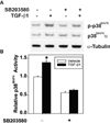
Figure 4. SB203580 blocked TGF-β-induced p38MAPK activity
RIE/GRPR cells were pretreated with or without SB203580 (10 µM) for 30 min followed by treatment with or without TGF-β (40 pM) for 60 min. A. Whole cell lysates were extracted and subjected to Western blot. p-p38MAPK (phospho-p38MAPK) and p38MAPKα proteins were measured using the specific antibodies, α-tubulin as a loading control. B. Cell lysates were assayed for p38MAPK activity using a recombinant MAPK-activated protein (MAPKAP) kinase-2 as a p38MAPK substrate. Results from duplicate wells were expressed as mean±SEM. *p < 0.05 compared with the group of vehicle control.
Figure 5
Figure 5. SB203580 attenuated TGF-β-induced apoptosis
The cells were seeded in 12-well plates for 24 h, then treated for 24 h with TGF-β1 (40 pM), BBS (100 nM), or TGF-β (40 pM) and BBS (100 nM) together, with or without pretreatment of SB203580 (10 µM) for 30 min. DNA fragmentation was quantified by a cell death detection assay. Results from triplicate wells are expressed as mean±SEM. *p < 0.05 compared with the group of vehicle control, † p < 0.05 compared with TGF-β alone, # p < 0.05 compared with the group with both TGF-β and BBS and without SB203580 pretreatment.

Similar articles

Cited by

References

    1. Babyatsky MW, Podolsky DK. Growth and development in the gastrointestinal tract. In: Yamada T, Alpers DH, Owyang C, Powell DW, editors. Text book of Gastroenterology. Philadelphia: JB Lippincott Co.; 1991. pp. 475–501.
    1. Ko TC, Bresnahan WA, Thompson EA. Intestinal cell cycle regulation. In: Meijer L, Guidet S, Philippe M, editors. Progress in Cell Cycle Research. New York: Plenum; 1997. pp. 43–52. - PubMed
    1. Cairnie AB, Lamerton LF, Steel GG. Cell proliferation studies in the intestinal epithelium of the rat. I. Determination of the kinetic parameters. Exp Cell Res. 1965;39:528–538. - PubMed
    1. Potten CS, Wilson JW, Booth C. Regulation and significance of apoptosis in the stem cells of the gastrointestinal epithelium. Stem Cells. 1997;15:82–93. - PubMed
    1. Hahm KB, Im YH, Parks TW, Park SH, Markowitz S, Jung HY, Green J, Kim SJ. Loss of transforming growth factor beta signalling in the intestine contributes to tissue injury in inflammatory bowel disease. Gut. 2001;49:190–198. - PMC - PubMed

Publication types

Substances