Review
. 2010 Feb;1803(2):164-73.
doi: 10.1016/j.bbamcr.2009.07.006. Epub 2009 Jul 23.

Formins and microtubules

Affiliations
Review

Formins and microtubules

F Bartolini et al. Biochim Biophys Acta. 2010 Feb.

Abstract

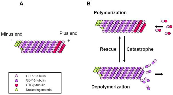
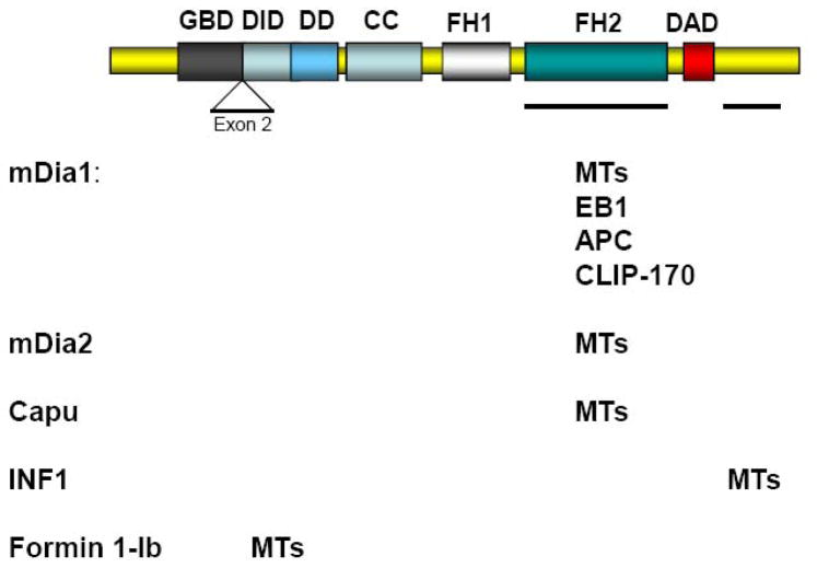
Formins have recently been recognized as prominent regulators of the microtubule (MT) cytoskeleton where they modulate the dynamics of selected MTs in interphase and mitosis. The association of formins with the MT cytoskeleton and their action on MT dynamics are relatively unexplored areas, yet growing evidence supports a direct role in their regulation of MT stability independent of their activity on actin. Formins regulate MT stability alone or in combination with accessory MT binding proteins that have previously been implicated in the stabilization of MTs downstream of polarity cues. As actin and MT arrays are typically remodeled downstream of signaling pathways that orchestrate cell shape and division, formins are emerging as excellent candidates for coordinating the responses of the cytoskeletal in diverse regulated and homeostatic processes.


Figures

Figure 1
Figure 1. Microtubule structure and dynamic instability
(A) A MT is a polarized polymer of α/β tubulin heterodimers. During polymerization the GTP bound to the β tubulin subunit is hydrolyzed (GTP is also bound to the α-subunit, but this is not exchangeable or hydrolyzed). GTP-bound subunits decorate the plus end while GDP-bound subunits form the core of the MT. The minus end of the MTs is usually anchored to nucleating material in cells and does not exchange subunits. (B) MT dynamic instability. In most cells MTs alternate between phases of polymerization and depolymerization at their plus ends. Transitions from shrinkage to growth are known as rescues while the opposite reaction is know as catastrophe. Most MT dynamics in vitro and in vivo can be described by the rates of growth and shrinkage and the frequencies of rescues and catastrophes.
Figure 2
Figure 2. MT binding regions on formins
The diagram represents the linear organization of domains in an idealized diaphanous-related formin. The domains known to interact with MTs or MT-interacting proteins for each of the formins are indicated. Note that most formins function as dimers and that Formin 1-Ib does not have a GBD or a DAD domain. GBD, GTPase binding domain; DID, DAD-interacting domain; DD, dimerization domain; CC, coiled-coil domain; FH1, formin homology 1; FH2, formin homology 2 domain; DAD, diaphanous autoregulatory domain.
Figure 3
Figure 3. Active mDia induces the formation of Glu MTs in fibroblasts
a–f, Constitutively actin ΔGBDmDia2–MycGFP was microinjected into nuclei of serum-starved NIH3T3 cells, and after 3–4 h, cells were fixed and immunostained for stable (Glu) and dynamic (Tyr) MTs and actin. One cell is shown in a–c, another in d–f (arrows denote expressing cells; GFP fluorescence is not shown for cell in d–f). a, GFP; b, e, Glu MTs; c, f, Tyr MTs; d, actin. The Glu MTs in the mDia2-expressing cell in (e) are oriented towards the wound edge, whereas those in the expressing cell in (b) are not. g–l, DAD-mycGFP was microinjected into nuclei of serum-starved NIH3T3 cells, and after 3 h cells were fixed and immunostained for Glu and Tyr MTs and actin. One cell is shown in g–j, another in k, l (arrows denote expressing cells; GFP fluorescence is not shown for cell in k, l). g, GFP; h, k, Glu MTs; i, l, Tyr MTs; j, actin. The Glu MTs in these cells were oriented towards the wound edge as seen in (e). Scale bars, 20 μm. Reproduced by permission of the authors [15].
Figure 4
Figure 4. Schematic indicating cellular sites of formin action on MT arrays
In migratory cells formins can regulate MTs at their plus ends (I), in proximity of the centrosome (II), or through binding along MTs (III). In cell division formins act on MT plus ends at kinetochore during mitosis (IV) and may act on central spindle MTs during cytokinesis (V).

Similar articles

Cited by

References

    1. Downing KH, Nogales E. Tubulin and microtubule structure. Curr Opin Cell Biol. 1998;10(1):16–22. - PubMed
    1. Downing KH, Nogales E. New insights into microtubule structure and function from the atomic model of tubulin. Eur Biophys J. 1998;27(5):431–6. - PubMed
    1. Nogales E, Wolf SG, Downing KH. Structure of the alpha beta tubulin dimer by electron crystallography. Nature. 1998;391(6663):199–203. - PubMed
    1. Nogales E, et al. High-resolution model of the microtubule. Cell. 1999;96(1):79–88. - PubMed
    1. Mitchison T, Kirschner M. Dynamic instability of microtubule growth. Nature. 1984;312(5991):237–42. - PubMed

MeSH terms