

Ventricular tachycardia from the healed myocardial infarction scar: validation of an animal model and utility of gene therapy
- PMID: 19631912
- PMCID: PMC2728453
- DOI: 10.1016/j.hrthm.2009.03.048
Ventricular tachycardia from the healed myocardial infarction scar: validation of an animal model and utility of gene therapy
Abstract
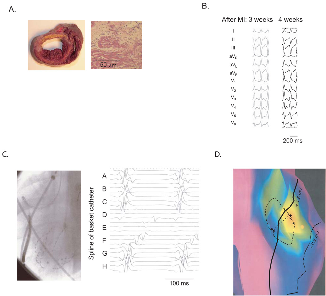
Life-threatening ventricular arrhythmias generally occur in the setting of structural heart disease. Current clinical options for patients at risk for these rhythm disturbances are limited. We developed a porcine model of inducible ventricular tachycardia originating in the border region of a healed myocardial infarction scar. After validating the model, we assessed gene transfer techniques, focusing on local modification of border zone tissues. We found that gene transfer of the dominant negative KCNH2-G628S mutation to the anteroseptal infarct border caused localized prolongation of effective refractory period in the target region and eliminated all ventricular arrhythmia inducibility. In this work, we characterize the animal model and review the gene transfer results.
Figures

Similar articles
-
Connexin43 gene transfer reduces ventricular tachycardia susceptibility after myocardial infarction.J Am Coll Cardiol. 2012 Sep 18;60(12):1103-10. doi: 10.1016/j.jacc.2012.04.042. Epub 2012 Aug 8. J Am Coll Cardiol. 2012. PMID: 22883636 Free PMC article.
-
Molecular ablation of ventricular tachycardia after myocardial infarction.Nat Med. 2006 Nov;12(11):1256-8. doi: 10.1038/nm1503. Epub 2006 Oct 29. Nat Med. 2006. PMID: 17072309 Free PMC article.
-
Antiarrhythmic and Anti-Inflammatory Effects of Sacubitril/Valsartan on Post-Myocardial Infarction Scar.Circ Arrhythm Electrophysiol. 2024 May;17(5):e012517. doi: 10.1161/CIRCEP.123.012517. Epub 2024 Apr 26. Circ Arrhythm Electrophysiol. 2024. PMID: 38666379
-
Catheter ablation of post myocardial infarction scar related ventricular tachycardia: role of entrainment.Indian Heart J. 2011 Jul-Aug;63(4):371-8. Indian Heart J. 2011. PMID: 22497054 Review. No abstract available.
-
Mechanism of Ventricular Tachycardia Occurring in Chronic Myocardial Infarction Scar.Circ Res. 2024 Feb 2;134(3):328-342. doi: 10.1161/CIRCRESAHA.123.321553. Epub 2024 Feb 1. Circ Res. 2024. PMID: 38300981 Free PMC article. Review.
Cited by
-
Assessment of distribution and evolution of mechanical dyssynchrony in a porcine model of myocardial infarction by cardiovascular magnetic resonance.J Cardiovasc Magn Reson. 2012 Jan 6;14(1):1. doi: 10.1186/1532-429X-14-1. J Cardiovasc Magn Reson. 2012. PMID: 22226320 Free PMC article.
-
Gene therapy to treat cardiac arrhythmias.Nat Rev Cardiol. 2015 Sep;12(9):531-46. doi: 10.1038/nrcardio.2015.61. Epub 2015 Apr 28. Nat Rev Cardiol. 2015. PMID: 25917154 Review.
-
Three-dimensional regional strain analysis in porcine myocardial infarction: a 3T magnetic resonance tagging study.J Cardiovasc Magn Reson. 2012 Dec 13;14(1):85. doi: 10.1186/1532-429X-14-85. J Cardiovasc Magn Reson. 2012. PMID: 23237210 Free PMC article.
-
Heterogeneous repolarization creates ventricular tachycardia circuits in healed myocardial infarction scar.Nat Commun. 2022 Feb 11;13(1):830. doi: 10.1038/s41467-022-28418-1. Nat Commun. 2022. PMID: 35149693 Free PMC article.
-
Development of a closed chest model of chronic myocardial infarction in Swine: magnetic resonance imaging and pathological evaluation.ISRN Cardiol. 2013 Oct 27;2013:781762. doi: 10.1155/2013/781762. eCollection 2013. ISRN Cardiol. 2013. PMID: 24282645 Free PMC article.
References
-
- Warnes CA, Roberts WC. Sudden coronary death: relation of amount and distribution of coronary narrowing at necropsy to previous symptoms of myocardial ischemia, left ventricular scarring and heart weight. Am J Cardiol. 1984;54:65–73. - PubMed
-
- Soo LH, Gray D, Hampton JR. Pathological features of witnessed out-of-hospital cardiac arrest presenting with ventricular fibrillation. Resuscitation. 2001;51:257–264. - PubMed
-
- Delacretaz E, Stevenson WG. Catheter ablation of ventricular tachycardia in patients with coronary heart disease - Part I: Mapping. Pacing Clin.Electrophysiol. 2001;24:1261–1277. - PubMed
-
- Pinto JMB, Boyden PA. Electrical remodeling in ischemia and infarction. Cardiovasc.Res. 1999;42:284–297. - PubMed
-
- Echt DS, Liebson PR, Mitchell LB, Peters RW, Obias-Manno D, Barker AH, Arensberg D, Baker A, Friedman L, Greene HL, et al. Mortality and morbidity in patients receiving encainide, flecainide, or placebo. The Cardiac Arrhythmia Suppression Trial. N Engl J Med. 1991;324:781–788. - PubMed
Publication types
MeSH terms
Grants and funding
LinkOut - more resources
Full Text Sources
Other Literature Sources
Medical
