

The role of transforming growth factor beta in cervical remodeling within the rat cervix
- PMID: 19631925
- PMCID: PMC2756469
- DOI: 10.1016/j.ajog.2009.06.002
The role of transforming growth factor beta in cervical remodeling within the rat cervix
Abstract
Objective: Transforming growth factor beta (TGFbeta) plays a central role in extracellular matrix remodeling. We hypothesized that TGFbeta signaling is involved in cervical remodeling. This study evaluated patterns within this signaling pathway.
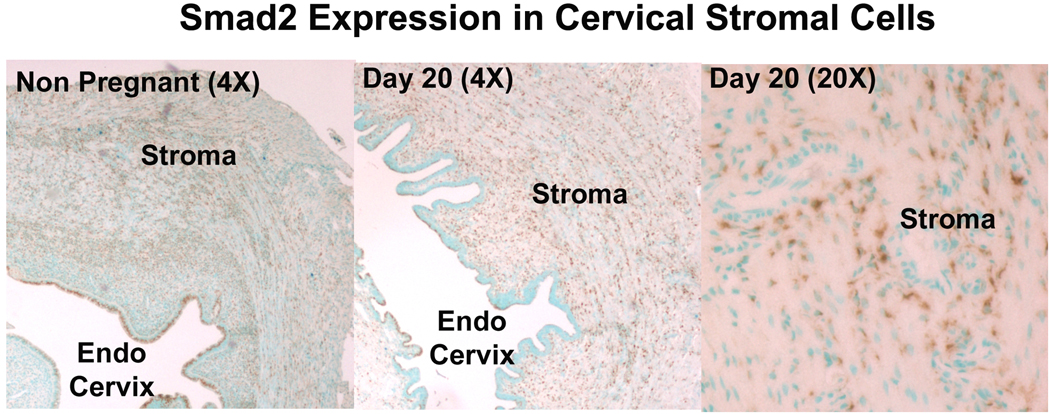
Study design: The cervices of nonpregnant and timed pregnant rats were obtained. Messenger ribonucleic acid (mRNA) expression of TGFbeta1, TGFbeta receptor 1 (TbetaR1), TbetaR2, and TbetaR3 was evaluated. Four animals were euthanized for each time point. Western blotting was performed for protein expression. Phosphorylated mothers against decapentaplegic (Smad)-2 and -3 phosphorylation was assessed to evaluate TGFbeta activation.
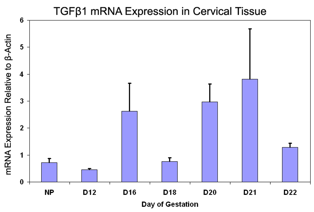
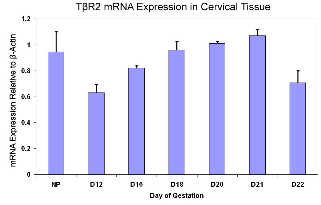
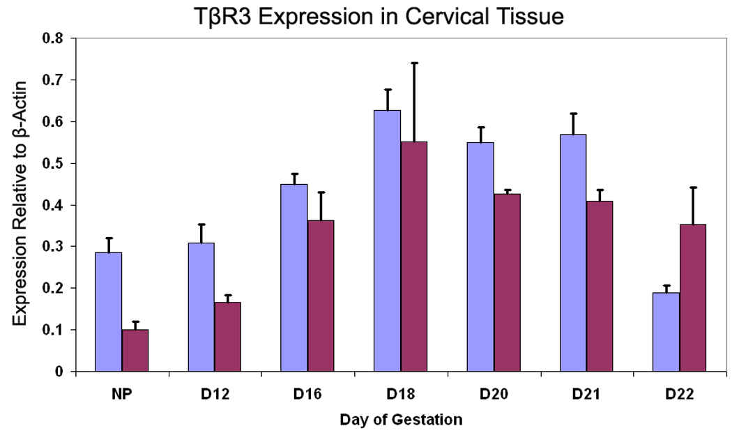
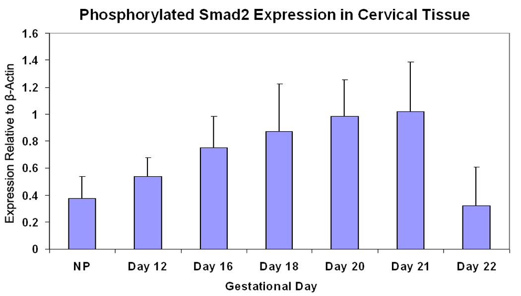
Results: TGFbeta1 mRNA increased through day 21 and declined on day 22 (analysis of variance, P = .001). TbetaR1 expression was unchanged. TbetaR2 and TbetaR3 mRNA expression was similar to TGFbeta1. TbetaR3 protein expression was similar to mRNA. Smad2 phosphorylation paralleled changes in TbetaR3.
Conclusion: Components of the TGFbeta signaling pathway increase during pregnancy along with Smad2 activation. The decline on day 22 correlates with a transition to the ripening phase supporting a role in cervical remodeling.
Figures
Similar articles
-
Knockdown of elF3a inhibits TGF‑β1‑induced extracellular matrix protein expression in keloid fibroblasts.Mol Med Rep. 2018 Mar;17(3):4057-4061. doi: 10.3892/mmr.2017.8365. Epub 2017 Dec 29. Mol Med Rep. 2018. PMID: 29286129
-
Oxidative stress is responsible for maternal diabetes-impaired transforming growth factor beta signaling in the developing mouse heart.Am J Obstet Gynecol. 2015 May;212(5):650.e1-11. doi: 10.1016/j.ajog.2015.01.014. Epub 2015 Jan 13. Am J Obstet Gynecol. 2015. PMID: 25595579 Free PMC article.
-
Transforming growth factor {beta}1 induces epithelial-mesenchymal transition by activating the JNK-Smad3 pathway in rat peritoneal mesothelial cells.Perit Dial Int. 2008 Jun;28 Suppl 3:S88-95. Perit Dial Int. 2008. PMID: 18552272
-
Nitric oxide induces TIMP-1 expression by activating the transforming growth factor beta-Smad signaling pathway.J Biol Chem. 2005 Nov 25;280(47):39403-16. doi: 10.1074/jbc.M504140200. Epub 2005 Sep 23. J Biol Chem. 2005. PMID: 16183640
-
A phosphatase controls the fate of receptor-regulated Smads.Cell. 2006 Jun 2;125(5):838-40. doi: 10.1016/j.cell.2006.05.015. Cell. 2006. PMID: 16751094 Review.
Cited by
-
The transcriptome of cervical ripening in human pregnancy before the onset of labor at term: identification of novel molecular functions involved in this process.J Matern Fetal Neonatal Med. 2009 Dec;22(12):1183-93. doi: 10.3109/14767050903353216. J Matern Fetal Neonatal Med. 2009. PMID: 19883264 Free PMC article.
References
-
- Goldenberg RL, Rouse DJ. Prevention of premature birth. N Engl J Med. 1998;339(5):313–320. - PubMed
-
- National Center for Health Statistics, final natality data. Retrieved February 4th, 2004, from www.marchofdimes.com/peristats.
-
- Mercer BM, et al. The Preterm Prediction Study: prediction of preterm premature rupture of membranes through clinical findings and ancillary testing. The National Institute of Child Health and Human Development Maternal-Fetal Medicine Units Network. Am J Obstet Gynecol. 2000;183(3):738–745. - PubMed
-
- Iams JD, et al. The length of the cervix and the risk of spontaneous premature delivery. National Institute of Child Health and Human Development Maternal Fetal Medicine Unit Network. N Engl J Med. 1996;334(9):567–572. - PubMed
Publication types
MeSH terms
Substances
Grants and funding
LinkOut - more resources
Full Text Sources
