

Down-regulation of multiple cell survival proteins in head and neck cancer cells by an apoptogenic mutant of adenovirus type 5
- PMID: 19631957
- PMCID: PMC2774264
- DOI: 10.1016/j.virol.2009.06.048
Down-regulation of multiple cell survival proteins in head and neck cancer cells by an apoptogenic mutant of adenovirus type 5
Abstract
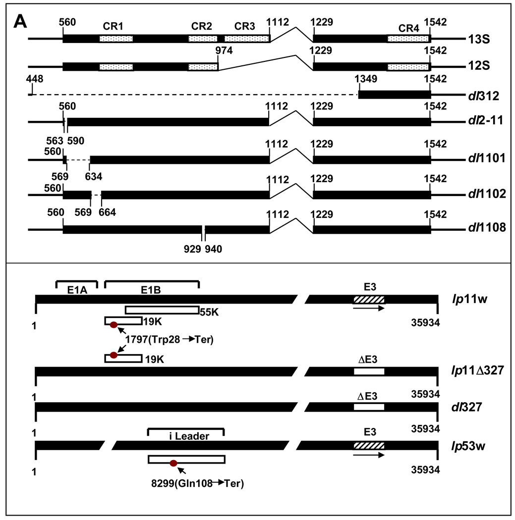
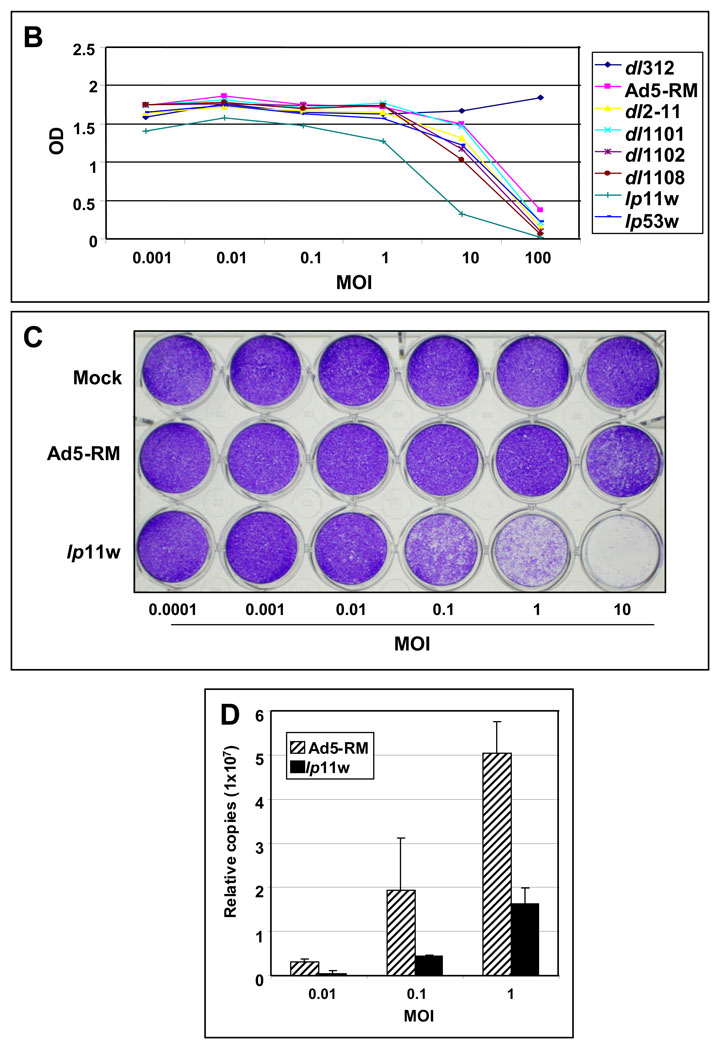
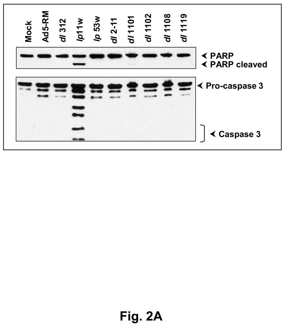
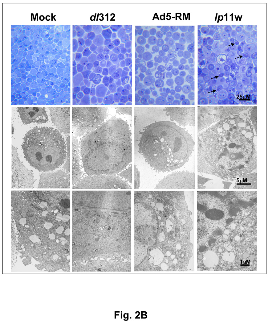
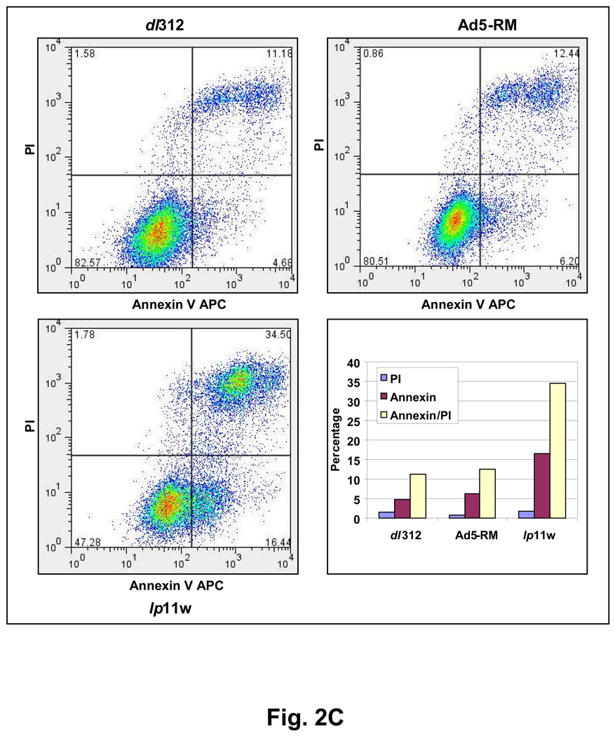
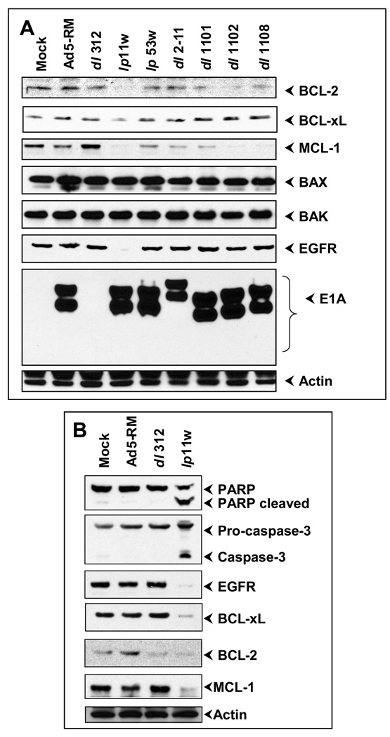
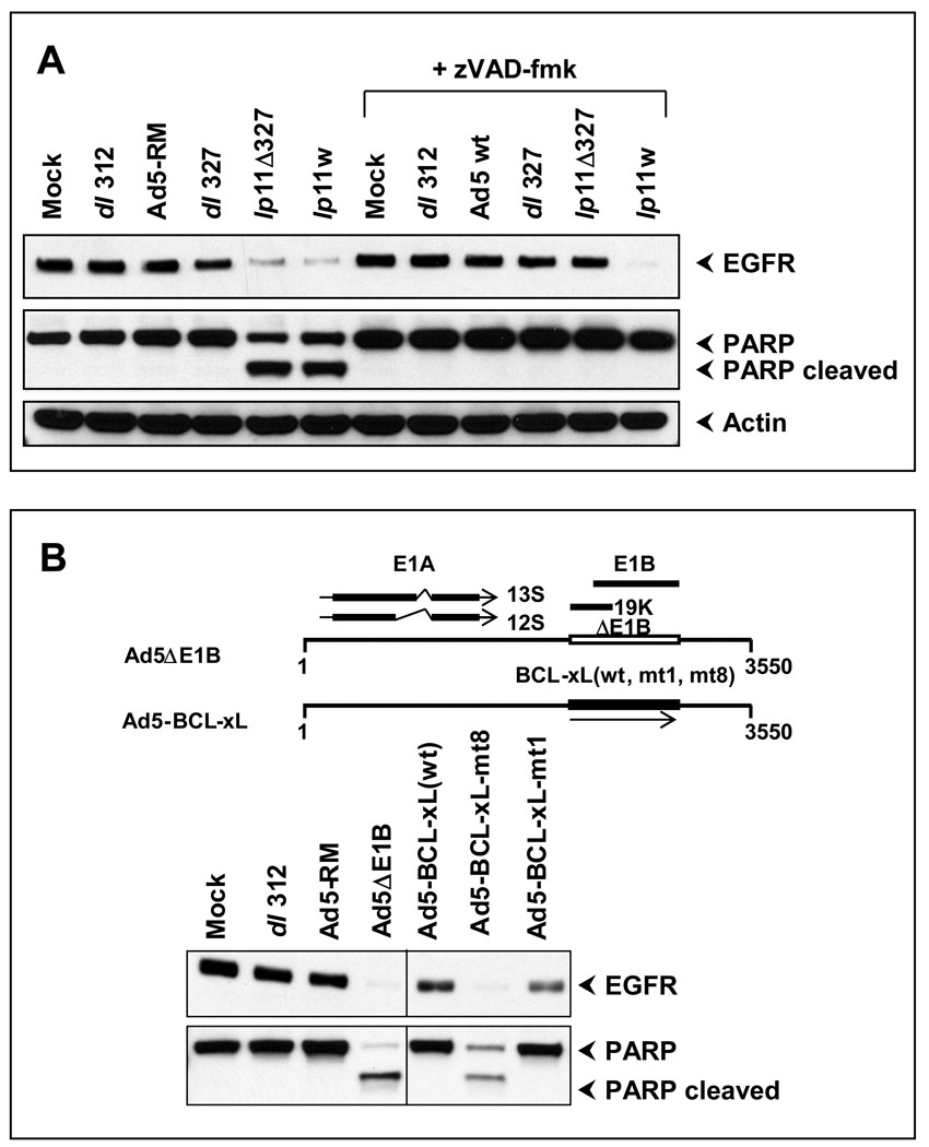
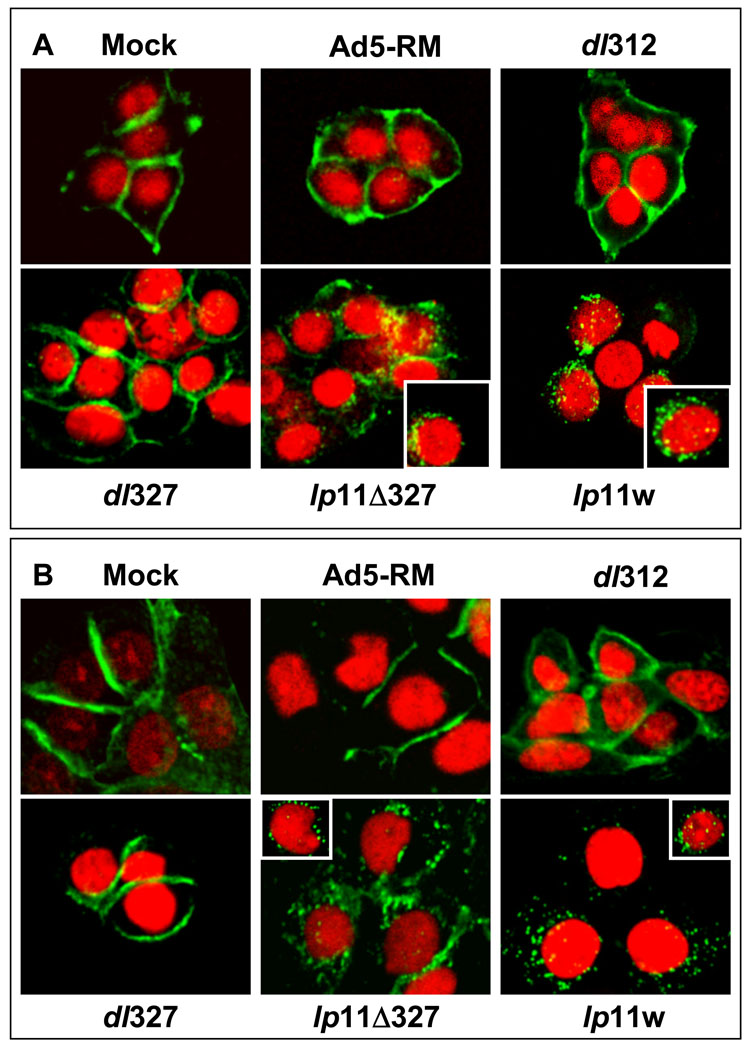
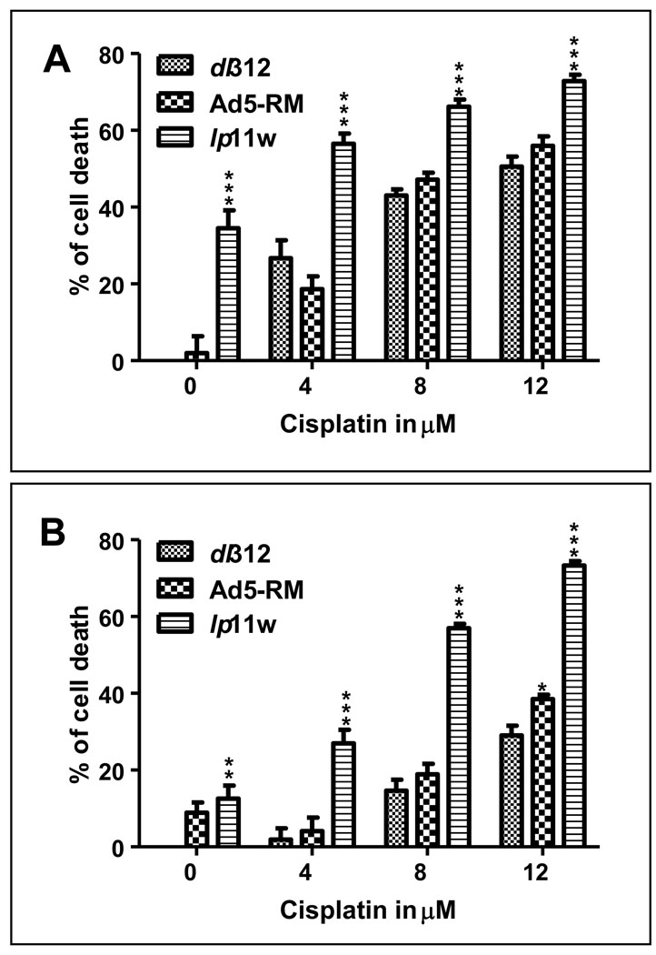
Head and neck squamous cell carcinomas (HNSCC) are one of the leading causes of cancer deaths world wide. Up-regulation of the epidermal growth factor receptor (EGFR) and BCL-2 family anti-apoptosis proteins in these cancers is linked to aggressive tumor growth, metastasis and chemoresistance. Infection of two HNSCC cell lines, SCC25 and CAL27 by an Ad5 mutant (lp11w) defective in coding for the viral anti-apoptosis protein, E1B-19K efficiently induced apoptotic cell death. In cells infected with lp11w there was a dramatic down-regulation of EGFR by apoptosis-dependent and -independent mechanisms. The levels of the anti-apoptotic proteins BCL-2, BCL-xL and MCL-1 were also down-regulated in lp11w-infected cells compared to uninfected or Ad5-RM infected cells. Infection with lp11w also enhanced sensitivity of the HNSCC cells to the chemotherapeutic drug cisplatin. Our results suggest that adenoviral vectors defective in E1B-19K would be valuable for efficient down-regulation of cell survival proteins and EGFR in epithelial cancers and could be exploited as oncolytic agents to treat HNSCCs.
Figures
Similar articles
-
Evaluation of apoptogenic adenovirus type 5 oncolytic vectors in a Syrian hamster head and neck cancer model.Cancer Gene Ther. 2014 Jun;21(6):228-237. doi: 10.1038/cgt.2014.22. Epub 2014 May 30. Cancer Gene Ther. 2014. PMID: 24874842 Free PMC article.
-
Hypertonicity counteracts MCL-1 and renders BCL-XL a synthetic lethal target in head and neck cancer.FEBS J. 2021 Mar;288(6):1822-1838. doi: 10.1111/febs.15492. Epub 2020 Aug 2. FEBS J. 2021. PMID: 32710568
-
Degradation of epidermal growth factor receptor mediates dasatinib-induced apoptosis in head and neck squamous cell carcinoma cells.Neoplasia. 2012 Jun;14(6):463-75. doi: 10.1596/neo.12300. Neoplasia. 2012. PMID: 22787428 Free PMC article.
-
The proteasome inhibitor MG132 potentiates TRAIL receptor agonist-induced apoptosis by stabilizing tBid and Bik in human head and neck squamous cell carcinoma cells.Exp Cell Res. 2012 Aug 1;318(13):1564-76. doi: 10.1016/j.yexcr.2012.04.003. Epub 2012 Apr 10. Exp Cell Res. 2012. PMID: 22513214
-
Antisense-mediated downregulation of anti-apoptotic proteins induces apoptosis and sensitizes head and neck squamous cell carcinoma cells to chemotherapy.Cancer Biol Ther. 2005 Jul;4(7):720-7. doi: 10.4161/cbt.4.7.1783. Epub 2005 Jul 2. Cancer Biol Ther. 2005. PMID: 15917659
Cited by
-
Safety of talimogene laherparepvec: a real-world retrospective pharmacovigilance study based on FDA Adverse Event Reporting System (FAERS).J Pharm Health Care Sci. 2024 Dec 18;10(1):79. doi: 10.1186/s40780-024-00388-0. J Pharm Health Care Sci. 2024. PMID: 39696696 Free PMC article.
-
Oncolytic Viruses for Cancer Therapy: Overcoming the Obstacles.Viruses. 2010 Jan;2(1):78-106. doi: 10.3390/v2010078. Viruses. 2010. PMID: 20543907 Free PMC article.
-
From scourge to cure: tumour-selective viral pathogenesis as a new strategy against cancer.PLoS Pathog. 2014 Jan;10(1):e1003836. doi: 10.1371/journal.ppat.1003836. Epub 2014 Jan 16. PLoS Pathog. 2014. PMID: 24453963 Free PMC article. Review.
-
Cell-Surface Integrins and CAR Are Both Essential for Adenovirus Type 5 Transduction of Canine Cells of Lymphocytic Origin.PLoS One. 2017 Jan 9;12(1):e0169532. doi: 10.1371/journal.pone.0169532. eCollection 2017. PLoS One. 2017. PMID: 28068367 Free PMC article.
-
Evaluation of apoptogenic adenovirus type 5 oncolytic vectors in a Syrian hamster head and neck cancer model.Cancer Gene Ther. 2014 Jun;21(6):228-237. doi: 10.1038/cgt.2014.22. Epub 2014 May 30. Cancer Gene Ther. 2014. PMID: 24874842 Free PMC article.
References
-
- Anderson RD, Haskell RE, Xia H, Roessler BJ, Davidson BL. A simple method for the rapid generation of recombinant adenovirus vectors. Gene Ther. 2000;7(12):1034–1038. - PubMed
-
- Bae SS, Choi JH, Oh YS, Perry DK, Ryu SH, Suh PG. Proteolytic cleavage of epidermal growth factor receptor by caspases. FEBS Lett. 2001;491(1–2):16–20. - PubMed
-
- Baselga J, Schoffski P, Rojo F, Dumez H, Ramos FJ, Macarulla T, Cajal R, Kisker O, Van Oosterom A, Tabernero J. A phase I pharmacokinetic (PK) and molecular pharmacodynamic (PD) study of the combination of two anti-EGFR therapies, the monoclonal antibody (MAb) cetuximab (C) and the tyrosine kinase inhibitor (TKI) gefitinib (G), in patients (pts) with advanced colorectal (CRC), head and neck (HNC) and non-small cell lung cancer (NSCLC) Journal of Clinical Oncology. 2006;24(18):122s–122s.
-
- Bauer JA, Trask DK, Kumar B, Los G, Castro J, Lee JS, Chen J, Wang S, Bradford CR, Carey TE. Reversal of cisplatin resistance with a BH3 mimetic, (−)-gossypol, in head and neck cancer cells: role of wild-type p53 and Bcl-xL. Mol Cancer Ther. 2005;4(7):1096–1104. - PubMed
Publication types
MeSH terms
Substances
Grants and funding
LinkOut - more resources
Full Text Sources
Medical
Research Materials
Miscellaneous
