

Review
doi: 10.1016/j.sbi.2009.07.003.
Epub 2009 Jul 23.
Peptides in the treatment of AIDS
Affiliations
- PMID: 19632107
- PMCID: PMC2763535
- DOI: 10.1016/j.sbi.2009.07.003
Item in Clipboard
Review
Peptides in the treatment of AIDS
Curr Opin Struct Biol.
2009 Aug.
Abstract
Fusion of HIV-1 and target cells is mediated by the envelope protein gp41 that undergoes a series of conformational changes during the process of infection. Knowledge of the structural biology of gp41 allows the design of potent peptide inhibitors that prevent the virus from entering lymphocytes and macrophages. The design of such inhibitors is the subject of this review.
Figures
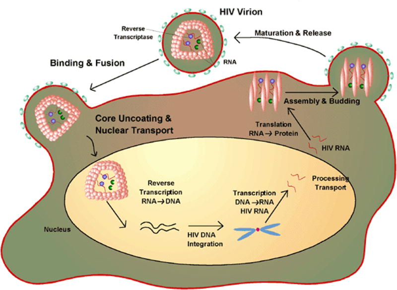
The life-cycle of HIV-1. The virus attaches to its target cell and subsequent to membrane fusion the RNA of the virus is reverse transcribed to DNA which is integrated into the cell’s genome. During it’s virulent phase the viral DNA is transcribed back to RNA which codes for various proteins required for virion assembly. Drugs can inhibit various events in the life cycle. The focus of the review is peptides that inhibit fusion to the target cell. Figure from http://www.wiley.com/legacy/college/boyer/0470003790/cutting_edge/aids_therapies/hiv_lifecycle.gif
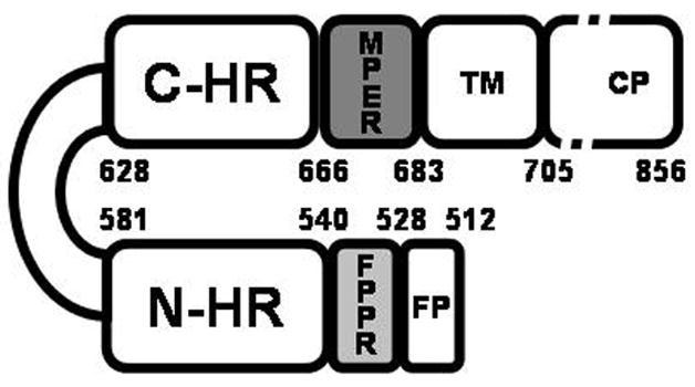
Schematic representation of HIV-1 gp41. The abbreviations represent the fusion peptide (FP); the fusion peptide proximal domain (FPPR); the N-terminal helical region (N-HR), the immunodominant loop region, the C-terminal helical region (C-HR), the membrane proximal region (MPER) and the transmembrane region (TM). The protein is numbered according to the HXB2 HIV-1 strain. The figure was adapted from [23].
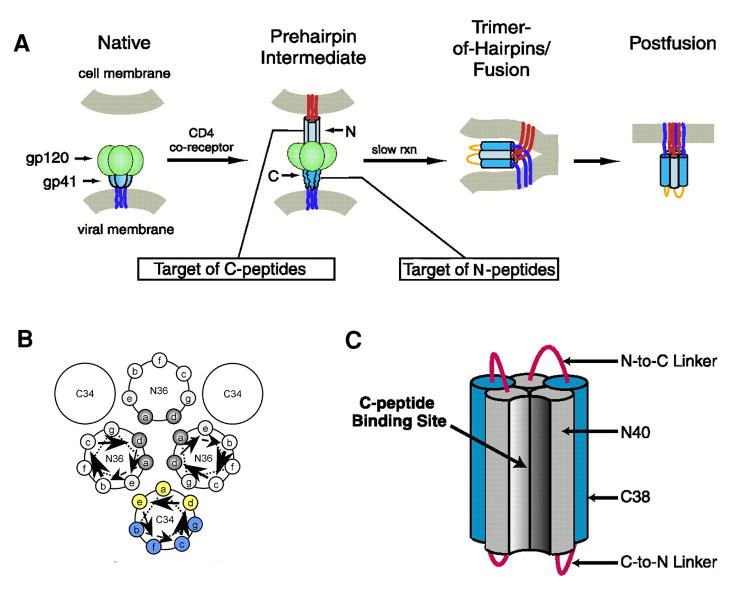
Mechanism of action of HIV-1 entry inhibitors. Panel A illustrates the involvement of gp41 in the fusion of the cell and viral membranes and the interference of peptide fusion inhibitors with this event. The inhibitors are believed to prevent formation of the 6-helix hairpin bundle necessary for juxtaposing the cell and viral membranes. C-HR peptides like T-20 bind to the N-HR trimeric core and N-peptides form a trimer which binds to the C-HR helix forming heterocomplexes that are defective in fusion. Panel B Schematic helical wheel diagram used in the design of C-peptides with increased helicity and water solubility. The positions that pack against the N-trimeric core (indicated in yellow) are less favored for replacement than those facing away (indicated in blue). Panel C. 5- Helix inhibitor. The hetero bundle lacks one C-peptide and thus provides a binding pocket for the C-HR peptide of gp41. Adapted from [13,38].
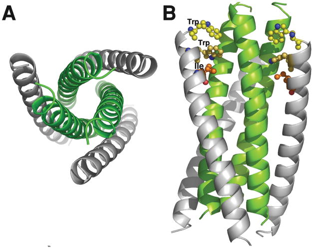
X-ray structure of the gp41 6-helix bundle. The bundle was formed from synthetic N36 and C34 peptides (see Table 1). Panel A shows an end view of the 6-HB with the C-peptides in grey and the trimeric core in green. Panel B is a side view illustrating the trimeric N-core interacting with the C-peptides. The pocket binding domain (PDB; WMEWDREI) residues Trp, Trp and Ile of C34 are shown explicitly. These interact with the hydrophobic pocket of the N-trimeric core. The model was built using coordinates from the PDB 1AIK.
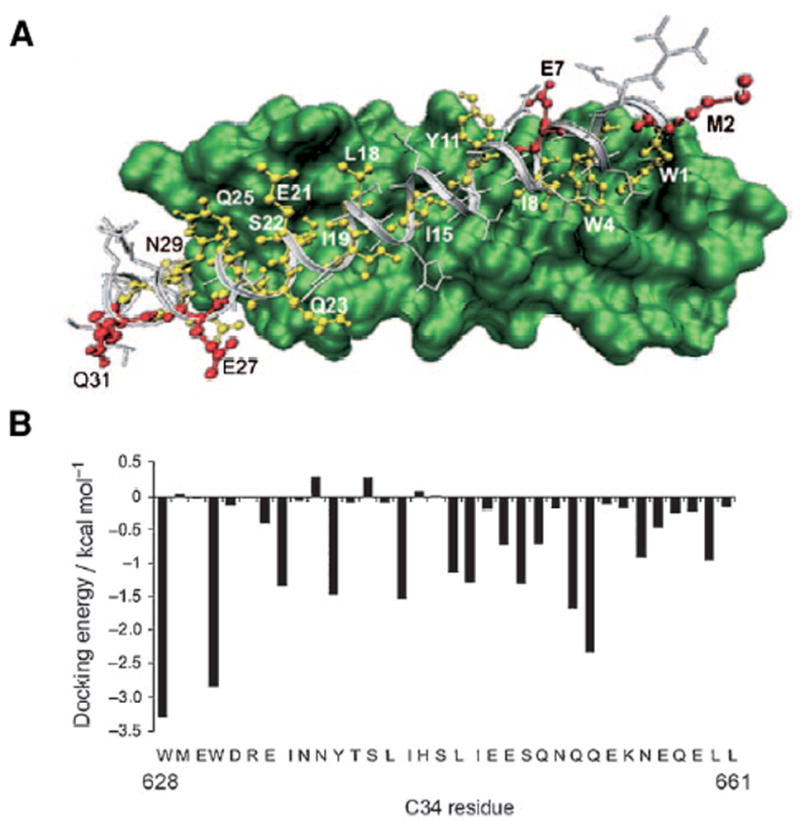
Docking of C34 into the N-trimeric core was used to choose residues for replacement by D-residues. The side chains indicated in red face away from the core and can be replaced with amino acids of opposite chirality. The docking energies provide additional guidance for positions that do not contribute to the binding affinity. Based on this structure C34M3 had D-residues inserted at positions 2, 27 and 31 and had potency comparable to C34 but was more soluble and more stable to protease digestion. Figure from [46].
Similar articles
-
Development of HIV-1 fusion inhibitors targeting gp41.Curr Med Chem. 2014 Jun;21(17):1976-96. doi: 10.2174/0929867321666131218094559. Curr Med Chem. 2014. PMID: 24350848 Review.
-
Peptide and non-peptide HIV fusion inhibitors.Curr Pharm Des. 2002;8(8):563-80. doi: 10.2174/1381612024607180. Curr Pharm Des. 2002. PMID: 11945159 Review.
-
Novel therapies based on mechanisms of HIV-1 cell entry.N Engl J Med. 2003 May 29;348(22):2228-38. doi: 10.1056/NEJMra022812. N Engl J Med. 2003. PMID: 12773651 Review. No abstract available.
-
Development of small molecule HIV-1 fusion inhibitors: linking biology to chemistry.Curr Pharm Des. 2013;19(10):1827-34. doi: 10.2174/1381612811319100007. Curr Pharm Des. 2013. PMID: 23092276 Review.
-
Qadirvirtide.Pak J Pharm Sci. 2011 Oct;24(4):593-5. Pak J Pharm Sci. 2011. PMID: 21959827 Review.
Cited by
-
Biophysical property and broad anti-HIV activity of albuvirtide, a 3-maleimimidopropionic acid-modified peptide fusion inhibitor.PLoS One. 2012;7(3):e32599. doi: 10.1371/journal.pone.0032599. Epub 2012 Mar 5. PLoS One. 2012. PMID: 22403678 Free PMC article.
-
Prediction of the interaction of HIV-1 integrase and its dicaffeoylquinic acid inhibitor through molecular modeling approach.Ethn Dis. 2010 Winter;20(1 Suppl 1):S1-45-9. Ethn Dis. 2010. PMID: 20521384 Free PMC article.
-
Design, synthesis, and evaluation of indole compounds as novel inhibitors targeting Gp41.Bioorg Med Chem Lett. 2010 Mar 1;20(5):1500-3. doi: 10.1016/j.bmcl.2010.01.111. Epub 2010 Jan 25. Bioorg Med Chem Lett. 2010. PMID: 20153190 Free PMC article.
-
A targeted covalent small molecule inhibitor of HIV-1 fusion.Chem Commun (Camb). 2021 May 6;57(37):4528-4531. doi: 10.1039/d1cc01013a. Chem Commun (Camb). 2021. PMID: 33956029 Free PMC article.
-
Anti-antimicrobial peptides: folding-mediated host defense antagonists.J Biol Chem. 2013 Jul 12;288(28):20162-72. doi: 10.1074/jbc.M113.459560. Epub 2013 Jun 4. J Biol Chem. 2013. PMID: 23737519 Free PMC article.
References
-
- Liu S, Wu S, Jiang S. HIV entry inhibitors targeting gp41: from polypeptides to small-molecule compounds. Curr Pharm Des. 2007;13:143–162. Comprehensive review of the design of HIV-1 inhibitors targeting gp41 (133 references) - PubMed
-
- Chan DC, Fass D, Berger JM, Kim PS. Core structure of gp41 from the HIV envelope glycoprotein. Cell. 1997;89:263–273. Determination of the crystal structure of the gp41 core formed using synthetic peptides. - PubMed
-
- Weissenhorn W, Dessen A, Harrison SC, Skehel JJ, Wiley DC. Atomic structure of the ectodomain from HIV-1 gp41. Nature. 1997;387:426–430. Description of the crystal structure of the gp41 core using crystals formed from biosynthetic and synthetic peptides. - PubMed
-
- Chan DC, Kim PS. HIV entry and its inhibition. Cell. 1998;93:681–684. - PubMed
Publication types
MeSH terms
Substances
Grants and funding
LinkOut - more resources
Full Text Sources
Other Literature Sources
Medical
