. 2009 Jul 23;138(2):286-99.
doi: 10.1016/j.cell.2009.05.045.

CD47 is an adverse prognostic factor and therapeutic antibody target on human acute myeloid leukemia stem cells

Affiliations

CD47 is an adverse prognostic factor and therapeutic antibody target on human acute myeloid leukemia stem cells

Ravindra Majeti et al. Cell. .

Abstract

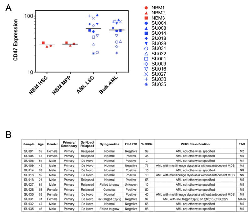
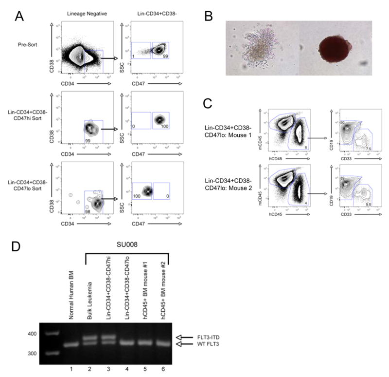
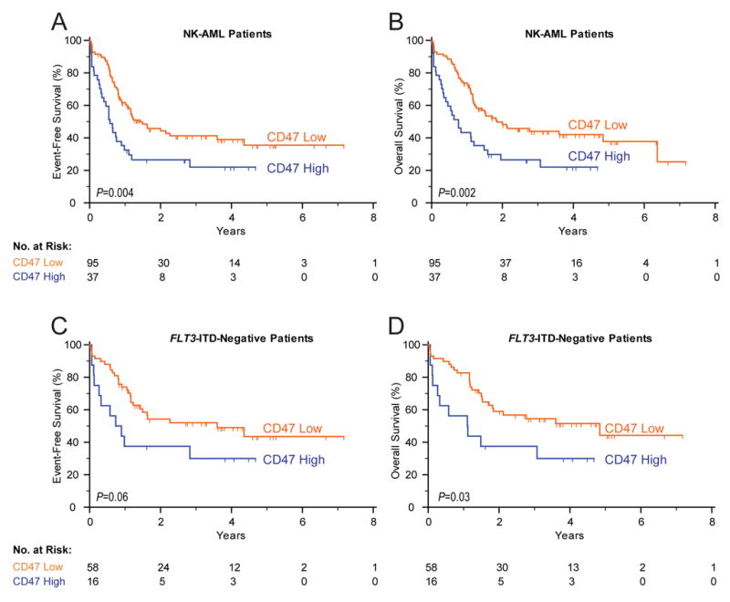
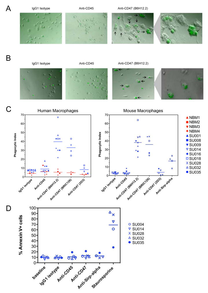
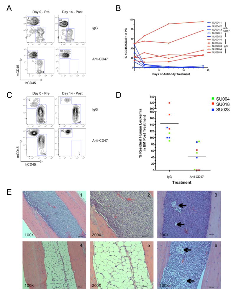
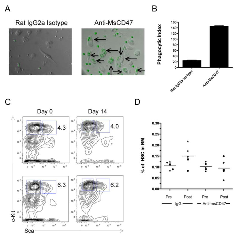
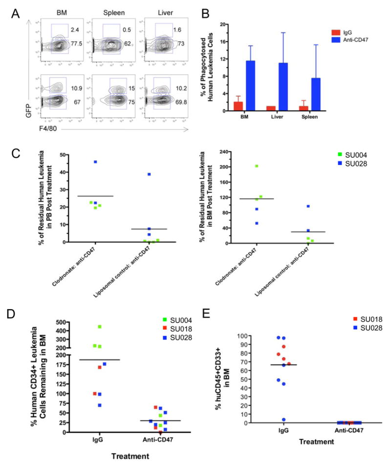
Acute myeloid leukemia (AML) is organized as a cellular hierarchy initiated and maintained by a subset of self-renewing leukemia stem cells (LSC). We hypothesized that increased CD47 expression on human AML LSC contributes to pathogenesis by inhibiting their phagocytosis through the interaction of CD47 with an inhibitory receptor on phagocytes. We found that CD47 was more highly expressed on AML LSC than their normal counterparts, and that increased CD47 expression predicted worse overall survival in three independent cohorts of adult AML patients. Furthermore, blocking monoclonal antibodies directed against CD47 preferentially enabled phagocytosis of AML LSC and inhibited their engraftment in vivo. Finally, treatment of human AML LSC-engrafted mice with anti-CD47 antibody depleted AML and targeted AML LSC. In summary, increased CD47 expression is an independent, poor prognostic factor that can be targeted on human AML stem cells with blocking monoclonal antibodies capable of enabling phagocytosis of LSC.


Figures

Figure 1
Figure 1. CD47 is More Highly Expressed on AML LSC Compared to Their Normal Counterparts
A. Relative CD47 expression on normal bone marrow HSC (Lin−CD34+CD38−CD90+) and MPP (Lin−CD34+CD38−CD90−CD45RA−), as well as LSC (Lin−CD34+CD38−CD90−) and bulk leukemia cells from human AML samples, was determined by flow cytometry. Mean fluorescence intensity was normalized for cell size and against lineage positive cells to account for analysis on different days. The same sample of normal bone marrow (red, n=3) or AML (blue, n=13) is indicated by the same symbol in the different populations. Normalized mean expression (and range) for each population was: HSC 30.6 (28.8–33.4), MPP 31.8 (30.0–33.4), LSC 59.8 (21.6–104.7), and bulk AML 56.3 (22.1–85.1). The differences between the mean expression of HSC with LSC (p=0.003), HSC with bulk leukemia (p=0.001), MPP with LSC (p=0.004), and MPP with bulk leukemia (p=0.002) were statistically significant using a 2-sided Student’s t-test. The difference between the mean expression of AML LSC compared to bulk AML was not statistically significant with p=0.50 using a paired 2-sided Student’s t-test. B. Clinical and molecular characteristics of primary human AML samples manipulated in vitro and/or in vivo.
Figure 2
Figure 2. Identification and Separation of Normal HSC From Leukemia Cells in the Same Patient Based On Differential CD47 Expression
A. CD47 expression on the Lin−CD34+CD38− LSC-enriched fraction of specimen SU008 was determined by flow cytometry. CD47hi- and CD47lo-expressing cells were identified and purified using FACS. The left panels are gated on lineage negative cells, while the right panels are gated on Lin−CD34+CD38− cells. B. Lin−CD34+CD38−CD47lo and Lin−CD34+CD38−CD47hi cells were plated into complete methylcellulose, capable of supporting the growth of all myeloid colonies. 14 days later, myeloid colony formation was determined by morphologic assessment. Representative CFU-G/M (left) and BFU-E (right) are presented. C. Lin−CD34+CD38−CD47lo cells were transplanted into 2 newborn NOG mice. 12 weeks later, the mice were sacrificed and the bone marrow was analyzed for the presence of human CD45+CD33+ myeloid cells and human CD45+CD19+ lymphoid cells by flow cytometry. D. Normal bone marrow HSC, bulk SU008 leukemia cells, Lin−CD34+CD38−CD47hi cells, Lin−CD34+CD38−CD47lo cells, or human CD45+ cells purified from the bone marrow of mice engrafted with Lin−CD34+CD38−CD47lo cells were assessed for the presence of the FLT3-ITD mutation by PCR. The wild type FLT3 and the FLT3-ITD products are indicated.
Figure 3
Figure 3. Increased CD47 Expression in Human AML is Associated with Poor Clinical Outcomes
Event-free (A,C) and overall (B,D) survival of 132 AML patients with normal cytogenetics (A,B) and the subset of 74 patients without the FLT3-ITD mutation (C,D). Patients were stratified into low CD47 and high CD47 expression groups based on an optimal threshold (28% high, 72% low) determined by microarray analysis from an independent training data set. The significance measures are based on log-likelihood estimates of the p-value, when treating the model with CD47 expression as a binary classification.
Figure 4
Figure 4. Monoclonal Antibodies Directed Against Human CD47 Preferentially Enable Phagocytosis of Human AML LSC by Human and Mouse Macrophages In Vitro
A,B. CFSE-labeled AML LSC were incubated with human peripheral blood-derived macrophages (A) or mouse bone marrow-derived macrophages (B) in the presence of IgG1 isotype control, anti-CD45 IgG1, or anti-CD47 (B6H12.2) IgG1 antibody. These cells were assessed by immunofluorescence microscopy for the presence of fluorescently-labeled LSC within the macrophages (indicated by arrows). C. CFSE-labeled AML LSC or normal bone marrow CD34+ cells were incubated with human (left) or mouse (right) macrophages in the presence of the indicated antibodies and then assessed for phagocytosis by immunofluorescence microscopy. The phagocytic index was determined for each condition by calculating the number of ingested cells per 100 macrophages. For AML LSC, the differences between isotype or anti-CD45 antibody with blocking anti-CD47 antibody treatment (B6H12.2 and BRIC126) were statistically significant with p<0.001 for all pairwise comparisons with human and mouse macrophages. For human macrophages, the differences between AML LSC and normal CD34+ cells were statistically significant for B6H12.2 (p<0.001) and BRIC126 (p=0.002). For mouse macrophages, the difference between istotype control and anti-SIRPα antibody was statistically significant (p=0.02). D. AML LSC were incubated in the presence of the indicated antibodies or the staurosporine positive control as described above, but in the absence of macrophages. At the end of the incubation, apoptotic cells were identified by Annexin V staining as determined by flow cytometry. No statistically significant increase in apoptosis was detected with any of the antibodies.
Figure 5
Figure 5. A Monoclonal Antibody Directed Against Human CD47 Depletes AML In Vivo
A–D. Newborn NOG mice were transplanted with AML LSC, and 8–12 weeks later, peripheral blood (C,D) and bone marrow (E,F) were analyzed for baseline engraftment prior to treatment with anti-CD47 (B6H12.2) or control IgG antibody (Day 0). Mice were treated with daily 100 microgram intraperitoneal injections for 14 days, at the end of which, they were sacrificed and peripheral blood and bone marrow were analyzed for the percentage of human CD45+CD33+ leukemia. (A) Pre- and post-treatment human leukemic chimerism in the peripheral blood from representative anti-CD47 antibody and control IgG-treated mice as determined by flow cytometry. (B) Summary of human leukemic chimerism in the peripheral blood assessed on multiple days during the course of treatment demonstrated elimination of leukemia in anti-CD47 antibody treated mice compared to control IgG treatment (p=0.007). (C) Pre- and post-treatment human leukemic chimerism in the bone marrow from representative anti-CD47 antibody or control IgG-treated mice as determined by flow cytometry. (D) Summary of human leukemic chimerism in the bone marrow on day 14 relative to day 0 demonstrated a dramatic reduction in leukemic burden in anti-CD47 antibody treated mice compared to control IgG treatment (p=0.006). E. H&E sections of representative mouse bone marrow cavities from mice engrafted with SU004 AML LSC post-treatment with either control IgG (panels 1,2) or anti-CD47 antibody (panels 4,5). IgG-treated marrows were packed with monomorphic leukemic blasts, while anti-CD47-treated marrows were hypocellular, demonstrating elimination of the human leukemia. In some anti-CD47 antibody-treated mice that contained residual leukemia, macrophages were detected containing phagocytosed pyknotic cells (panels 3,6 arrows).
Figure 6
Figure 6. A Monoclonal Antibody Directed Against Mouse CD47 Enables Phagocytosis of Mouse AML and Does Not Deplete Normal HSC In Vivo
A. GFP+ mouse AML cells were incubated with mouse bone marrow-derived macrophages in vitro in the presence of 10 micrograms/ml of rat IgG2a isotype control or anti-mouse CD47 antibody for two hours. Phagocytosis of GFP+ leukemia cells was observed by fluorescence microscopy (arrows). B. Quantitative analysis of phagocytosis was determined by calculating the phagocytic index in triplicate assays. Anti-MsCD47 antibody enabled a statistically significant increase in phagocytosis of mouse leukemia cells compared to isotype control (p<0.001). C,D. C57BL/6 wild type mice were treated for 14 days with daily 200 microgram intraperitoneal injections of either anti-msCD47 or rat IgG control antibody. Bone marrow from these mice was aspirated pre- and post-treatment and indicated no effect of either treatment on the frequency of Lin−Kit+Sca+ (KLS) cells (C) or Lin−Kit+Sca+Flk2−CD34− HSC (D) in the bone marrow. Representative flow cytometry plots are shown in panel C. No differences in the percentage of HSC pre and post-treatment were observed with either control IgG (p=0.09) or anti-msCD47 (p=0.81).
Figure 7
Figure 7. A Monoclonal Antibody Directed Against Human CD47 Enables Phagocytosis of AML In Vivo and Targets LSC
A,B. Flow cytometry plots (A) and quantitation (B) from NOG mice engrafted with lentivirally-transduced GFP-positive SU028 AML LSC, 4 hours after treatment with a single 100 microgram intraperitoneal dose of anti-CD47 antibody (B6H12.2) or control IgG (n=2 for each). Cell suspensions from bone marrow, spleen, and liver were stained for human CD45 and mouse F4/80, which recognizes phagocytes. All plots are gated on human CD45-negative cells. Double positive events represent GFP-positive leukemia cells within mouse phagocytes. C. NOG mice engrafted with the indicated AML LSC, were treated with liposomal clodronate to deplete phagocytes, and administered daily intraperitoneal injections of anti-CD47 antibody for 14 days. The percentage of residual human leukemia cells in the peripheral blood (left) and bone marrow (right) was determined as described above. Depletion of phagocytes resulted in a statistically significant inhibition of the ability of anti-CD47 antibody to eliminate human AML from both the peripheral blood (p=0.03) and bone marrow (p=0.04). Clodronate treatment by itself had no effect on leukemic engraftment (Supplemental Figure 12 A,B). D. The percentage of human CD34+ LSC-enriched human leukemia cells remaining in the bone marrow after treatment with either IgG control or anti-CD47 antibody was determined by flow cytometry. Treatment with anti-CD47 antibody resulted in a statistically significant decrease (p<0.001) compared to the control. E. 500,000 whole bone marrow cells from IgG control (n=12) or anti-CD47 (n=9) antibody-treated mice were secondarily transplanted into NOG mice. 12 weeks later, secondary mice were sacrificed and analyzed for human leukemia engraftment in the peripheral blood (Supplemental Figure 13) and bone marrow as described above. Secondary mice transplanted from IgG treated mice engrafted human leukemia in the bone marrow, while secondary mice transplanted from anti-CD47 treated mice developed no engraftment (p<0.001). Statistical significance was determined using Fisher’s exact test.

Comment in

References

    1. Adams GP, Weiner LM. Monoclonal antibody therapy of cancer. Nat Biotechnol. 2005;23:1147–1157. - PubMed
    1. Ailles LE, Gerhard B, Hogge DE. Detection and characterization of primitive malignant and normal progenitors in patients with acute myelogenous leukemia using long-term coculture with supportive feeder layers and cytokines. Blood. 1997;90:2555–2564. - PubMed
    1. Barclay AN, Brown MH. The SIRP family of receptors and immune regulation. Nat Rev Immunol. 2006;6:457–464. - PubMed
    1. Blazar BR, Lindberg FP, Ingulli E, Panoskaltsis-Mortari A, Oldenborg PA, Iizuka K, Yokoyama WM, Taylor PA. CD47 (integrin-associated protein) engagement of dendritic cell and macrophage counterreceptors is required to prevent the clearance of donor lymphohematopoietic cells. J Exp Med. 2001;194:541–549. - PMC - PubMed
    1. Bonnet D, Dick JE. Human acute myeloid leukemia is organized as a hierarchy that originates from a primitive hematopoietic cell. Nat Med. 1997;3:730–737. - PubMed

Publication types