. 2009 Jul 23;138(2):377-88.
doi: 10.1016/j.cell.2009.05.022.

The tumor suppressor Par-4 activates an extrinsic pathway for apoptosis

Affiliations

The tumor suppressor Par-4 activates an extrinsic pathway for apoptosis

Ravshan Burikhanov et al. Cell. .

Erratum in

  • Cell. 2009 Sep 4;138(5):1032

Abstract

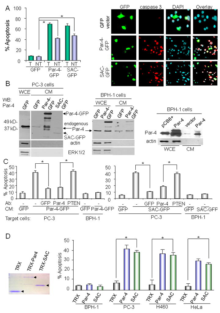
Prostate apoptosis response-4 (Par-4) is a proapoptotic protein with intracellular functions in the cytoplasm and nucleus. Unexpectedly, we noted Par-4 protein is spontaneously secreted by normal and cancer cells in culture, and by Par-4 transgenic mice that are resistant to spontaneous tumors. Short exposure to endoplasmic reticulum (ER) stress-inducing agents further increased cellular secretion of Par-4 by a brefeldin A-sensitive pathway. Secretion occurred independently of caspase activation and apoptosis. Interestingly, extracellular Par-4 induced apoptosis by binding to the stress response protein, glucose-regulated protein-78 (GRP78), expressed at the surface of cancer cells. The interaction of extracellular Par-4 and cell surface GRP78 led to apoptosis via ER stress and activation of the FADD/caspase-8/caspase-3 pathway. Moreover, apoptosis inducible by TRAIL, which also exerts cancer cell-specific effects, is dependent on extracellular Par-4 signaling via cell surface GRP78. Thus, Par-4 activates an extrinsic pathway involving cell surface GRP78 receptor for induction of apoptosis.


Figures

Figure 1
Figure 1. Par-4 and the SAC protein are secreted and induce apoptosis
A. Bystander effect of Par-4 and SAC domain. PC-3 cells were transiently transfected with the indicated expression constructs, and subjected to ICC for active caspase-3 to detect apoptosis, and counterstained with DAPI (right panel). Quantification of apoptosis in transfected (T) and non-transfected (NT) cells is presented (left panel). B. Conditioned medium (CM) from transfected cells contains secreted Par-4-GFP and SAC-GFP protein. The CM from cells transfected with the indicated expression constructs or pCB6+ vector was subjected to Western blot (WB) analysis with the Par-4, ERK1/2, or actin antibody. Appropriate whole-cell extracts (WCE) were used as controls. C. The secreted fusion proteins induce apoptosis in target PC-3 cells. Cells were treated with the CM from the indicated PC-3 transfectants (GFP, Par-4-GFP, or SAC-GFP) or with the CM that was pre-incubated for 30 min with neutralizing antibody (Ab) for GFP, Par-4, or PTEN. After 24h, the cells were scored for apoptosis by caspase-3 activation using ICC analysis. D. Recombinant Par-4 and SAC protein induce apoptosis in cancer cells. Recombinant protein was subjected to SDS-PAGE and Coomassie blue (CB) staining (left panel). Various cancer or non-transformed cells were treated with 100 nM amounts of each protein for 24 h (right panel). The cells were scored for apoptosis by ICC for active caspase-3. Panels C & D: Mean values (+ standard deviation bars) of three separate experiments are shown. Asterisk (*) indicates the difference is statistically significant (P < 0.001) by the Student’s t test.
Figure 2
Figure 2. Par-4 protein is secreted by an ER-stress inducible pathway
A. Par-4 protein is secreted by a BFA-sensitive pathway independently of apoptosis. Cells were left untreated (UT) or pre-treated with BFA (1 μM) for 30 min as indicated, then exposed to TRAIL (100 ng/ml) for 30, 60, and 180 min. Whole-cell extracts (WCE) or CM were prepared, and subjected to Western blot (WB) analysis for Par-4 or actin. The CM was also subjected to SDS-PAGE followed by Coomassie blue (CB) staining to reveal the 68 kDa BSA band for loading control (upper panels and lower left panel). PC-3 or PC-3/DN-FADD cells were also treated with TRAIL or left untreated (UT) for 24 h, and scored for apoptosis by ICC for active caspase 3 (lower right panel). Mean values (+ standard deviation bars) of three separate experiments are shown. Asterisk (*) indicates the difference is statistically significant (P < 0.001) by the Student’s t test. B. Par-4 protein is secreted by an ER-stress inducible pathway. PC-3 cells were left untreated (UT) or treated with thapsigargin (TG,100 nM) or tunicamycin (TU, 5 μM) in the presence or absence of BFA (1 μM) for the indicated time intervals. CM was prepared, and subjected to SDS-PAGE followed by staining with Coomassie blue (CB; upper panel). Also, whole-cell extracts from untreated (UT) PC-3 cells or PC-3 cells treated with vehicle, TG, TU, or TRAIL for various time intervals were subjected to Western blot (WB) analysis for Par-4, GRP78, and actin (middle panel), or CHOP/GADD153 and actin (lower panel).
Figure 3
Figure 3. Par-4, via its SAC domain, binds to GRP78 and co-localizes with GRP78 in the ER and at the cell membrane
A. Par-4 binds to GRP78. Whole-cell extracts from PC-3 cells were subjected to immunoprecipitation (IP) with antibodies for Par-4, GRP78, or GFP, and the immunoprecipitated complexes were resolved by SDS-PAGE and subjected to Western blot (WB) analysis for GRP78 and Par-4. PC-3 whole-cell extracts were used as input. B. Par-4 binds to GRP78 via its SAC domain. PC-3 cells were transfected with GFP, Par-4-GFP, or SAC-GFP expression constructs or left untransfected, and the cell lysates were subjected to immunoprecipitation (IP) with Par-4 or GFP antibody. The immunoprecipitated complexes were subjected to Western blot (WB) analysis for GRP78 or GFP. Whole-cell extracts from the transfectants were used as input. C. Recombinant Par-4, SAC, and TRAIL individually increase GRP78 expression at the cell surface. PC-3 cells were treated with TRAIL (100 ng/ml), TRX-Par-4 (100 nM), TRX-SAC (100 nM), or TRX (250 nM) for various time intervals. The cells were collected after 6 h (unfixed, so the antibody could detect only cell surface GRP78) and subjected to FACS analysis for GRP78. Mean values (+ standard deviation bars) of three separate experiments are shown. Asterisk (*) indicates the difference is statistically significant (P < 0.001) by the Student’s t test. D. GRP78 binds Par-4 at the cell surface. Cells were treated with TRAIL (100 ng/ml) or vehicle for 6 h, and then labeled with cell-impermeable sulfo-NHS-LC-biotin. Whole-cell extracts (WCE) of the TRAIL-treated/biotinylated samples were subjected to immunoprecipitation (IP) with the indicated antibodies (left panels). For the pull-down of biotinylated GRP78 from TRAIL-treated cells, the corresponding WCE (10 μg) were incubated with polyhistidine-tagged recombinant proteins TRX-Par-4 or TRX control (2 μg of each) for 2 h at 4°C. Bound complexes were purified with metal affinity resin beads (middle panels). The immunoprecipitated proteins (left panels), bound complexes (middle panels), and also the corresponding WCE (indicated as ‘input’, right panels) were subjected to SDS-PAGE, and blotted with avidin-HRP or with GRP78, Par-4, or TRX antibody. The ~50 kDa band indicated by the asterisk is unidentified. E. Par-4 co-localizes with GRP78 in the ER and in the cell membrane. Cells were treated with TRAIL (100 ng/ml) or vehicle for 6 h, and subjected to ICC for Par-4 (green fluorescence), GRP78 (red fluorescence), and nuclei were revealed by DAPI (cyan fluorescence). White arrows show co-localization of Par-4 and GRP78 in merged images (yellow fluorescence). A total of 400 cells were scored for co-localization of Par-4 and GRP78 at the cell membrane in each experiment, and mean values (+ standard deviation bars) of three separate experiments are shown in the right panel. Asterisk (*) indicates the difference is statistically significant (P < 0.0001) by the Student’s t test. Magnification: For co-localization images- PC-3/TRAIL (200x); PC-3/vehicle (120x); and BPH-1 (80x); for all other BPH-1 images (40x) and PC-3 images (80x).
Figure 4
Figure 4. Extracellular Par-4, SAC, and TRAIL are dependent on cell surface GRP78 for induction of apoptosis
A. Extracellular GRP78 neutralizes the apoptotic action of TRX-Par-4. PC-3 cells were pre-treated with vehicle, GST, or GRP78 protein, and then treated with TRX-Par-4, TRX protein, or vehicle. After 24 h, cells were subjected to ICC for active caspase-3 (green fluorescence, see Supplemental Figures S4B), using DAPI (pseudo-colored red, lower panel) to reveal nuclei, and apoptosis was quantified. The integrity of the purified proteins was verified by SDS-PAGE and Coomassie blue (CB) staining using protein molecular weight markers (upper panel). B. Neutralizing antibody (NT-GRP78) directed toward the N-terminal region of GRP78 inhibits apoptosis by extracellular Par-4. Cells were pre-incubated with the indicated antibodies for 30 minutes, and then treated with TRX-Par-4 or TRX (100 nM of each) for 24 h. Apoptotic cells were scored by ICC for active caspase-3. C. NT-GRP78-antibody neutralizes the apoptotic action of exogenous TRAIL, Par-4, and SAC. Cells were pre-treated with PTEN, NT-GRP78, DR4, or DR5 antibody for 30 min, and then treated with TRX-Par-4 (rPar-4), TRX-SAC (rSAC), or TRX. After 24 h, apoptosis was quantified by ICC for active caspase-3. D. Par-4- and NT-GRP78-antibodies individually neutralize the apoptotic action of TRAIL. PC-3 cells were pre-treated with PTEN, NT-GRP78, or Par-4 antibody, then treated with TRAIL (100 ng/ml) or vehicle. After 24 h, apoptosis was quantified by ICC for active caspase-3. E. Par-4, secreted in the serum of Par-4-transgenic mice, induces GRP78-mediated apoptosis in PC-3 cells. Bone marrow cells from littermate control, GFP-, and Par-4-GFP-transgenic mice were analyzed by ICC for GFP (upper left panel), and the serum from these mice was examined by Western blot for secreted Par-4 (upper right panel). Loading was verified by CB staining for serum albumin (upper right panel). Cells were treated with the serum samples (20% final concentration) either in the absence or presence of neutralizing antibodies, and, after 24 h, apoptosis was quantified by ICC for active caspase-3 (lower panel). In panels A-E, mean values (+ standard deviation bars) of three separate experiments are shown. Asterisk (*) indicates the difference is statistically significant (P < 0.001) by the Student’s t test.
Figure 5
Figure 5
Cell membrane-directed GRP78 overrides the effect of intracellular Par-4 knock-down and restores sensitivity to TRX-Par-4 A. Endogenous Par-4 expression is essential for apoptosis by extracellular Par-4. PC-3 cells were transfected with siRNA duplexes for Par-4 or control siRNA. The cells were treated with TRX-Par-4, TRX-SAC, or GFP protein for control for 24 h, and apoptotic cells were scored by evaluating active caspase-3 by ICC (upper panel). Western blot analysis (lower panel) confirmed both knock-down of Par-4 expression by siRNA and its effect on GRP78 expression. B. Intracellular Par-4 regulates trafficking of cell surface GRP78. PC-3 cells were transfected with siRNA duplexes for Par-4 or control siRNA, treated with TRX or TRX-Par-4, and processed as intact, unfixed cells for expression of cell surface GRP78 by FACS analysis (lower panel). Knock-down of intracellular Par-4 by siRNA was confirmed by Western blot analysis of corresponding whole-cell lysates (upper panel). C. Expression of membrane-targeted GRP78 restores sensitivity to TRX-Par-4 following intracellular Par-4 knock-down. PC-3 cells were transfected with siRNA duplexes for Par-4 or control siRNA, and knock-down of intracellular Par-4 was confirmed as indicated in Panel B. The cells were then transfected with vector, cell membrane-directed full-length GRP78 (mGRP78), or ΔN mutant of GRP78 (mΔN-GRP78), and expression of endogenous and ectopic GRP78 in whole-cell lysates was confirmed by Western blot analysis (left panel inset). Finally, the transfectants were subjected to treatment with TRX or TRX-Par-4 for 24 h, and apoptotic cells were scored by evaluating active caspase-3 by ICC (left panel). Expression of cell surface GRP78, as determined by FACS analysis, is shown for Par-4 knock-down cells transfected with the mGRP78 construct or vector and subsequently treated with TRX-Par-4 (right panel). In panels A-C, mean values (+ standard deviation bars) of three separate experiments are shown. Asterisk (*) indicates the difference is statistically significant (P < 0.001) by the Student’s t test.
Figure 6
Figure 6
Model for apoptosis by extracellular Par-4 ER stress (a), induced by extracellular insults such as TRAIL, causes translocation of the Par-4-GRP78 complex from the ER to the plasma membrane (b). This leads to elevated expression of GRP78 and Par-4 at the plasma membrane (c), and secretion of Par-4 (d). Extracellular Par-4 binds to cell surface GRP78 (e), and induces yet more ER stress (f), thereby activating the loop for translocation of ER GRP78 to the plasma membrane, in an intracellular Par-4-dependent manner. Finally, ER stress involves up-regulation of the ER component phospho-PERK, and activation of caspase-8 in a PERK-dependent manner (g). Activation of caspase-8 is also dependent on activation of FADD. Collectively, these events lead to activation of caspase-3 and apoptosis. This model is not restricted to prostate cancer cells, as the key features were recapitulated in lung and cervical cancer cells (Figure S11).

Comment in

Similar articles

Cited by

References

    1. Abdelrahim M, Newman K, Vanderlaag K, Samudio I, Safe S. 3,3′-diindolylmethane (DIM) and its derivatives induce apoptosis in pancreatic cancer cells through endoplasmic reticulum stress-dependent upregulation of DR5. Carcinogenesis. 2006;27:717–728. - PubMed
    1. Arap MA, Lahdenranta J, Mintz PJ, Hajitou A, Sarkis AS, Arap W, Pasqualini R. Cell surface expression of the stress response chaperone GRP78 enables tumor targeting by circulating ligands. Cancer Cell. 2004;6:275–284. - PubMed
    1. Chakraborty M, Qiu SG, Vasudevan KM, Rangnekar VM. Par-4 drives trafficking and activation of Fas and Fasl to induce prostate cancer cell apoptosis and tumor regression. Cancer Res. 2001;61:7255–7263. - PubMed
    1. Cheema SK, Mishra SK, Rangnekar VM, Tari AM, Kumar R, Lopez-Berestein G. Par-4 transcriptionally regulates Bcl-2 through a WT1-binding site on the bcl-2 promoter. J Biol Chem. 2003;278:19995–20005. - PubMed
    1. Cook J, Krishnan S, Ananth S, Sells SF, Shi Y, Walther MM, Linehan WM, Sukhatme VP, Weinstein MH, Rangnekar VM. Decreased expression of the pro-apoptotic protein Par-4 in renal cell carcinoma. Oncogene. 1999;18:1205–1208. - PubMed

Publication types