. 2009 Sep;49(19):2386-92.
doi: 10.1016/j.visres.2009.07.011. Epub 2009 Jul 24.

Relative peripheral hyperopic defocus alters central refractive development in infant monkeys

Affiliations

Relative peripheral hyperopic defocus alters central refractive development in infant monkeys

Earl L Smith 3rd et al. Vision Res. 2009 Sep.

Abstract

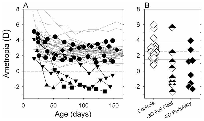
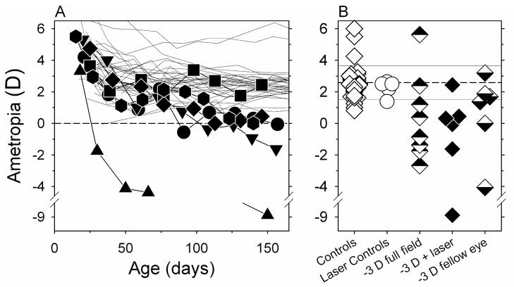
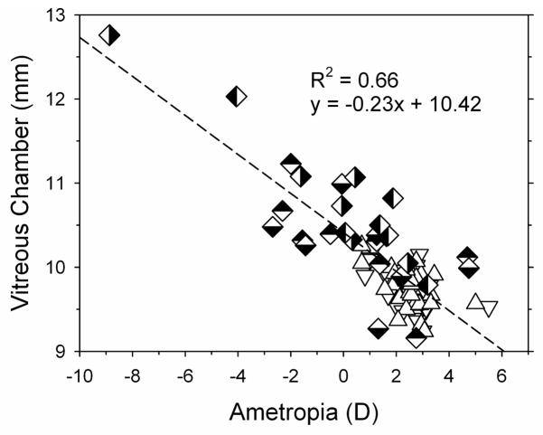
Understanding the role of peripheral defocus on central refractive development is critical because refractive errors can vary significantly with eccentricity and peripheral refractions have been implicated in the genesis of central refractive errors in humans. Two rearing strategies were used to determine whether peripheral hyperopia alters central refractive development in rhesus monkeys. In intact eyes, lens-induced relative peripheral hyperopia produced central axial myopia. Moreover, eliminating the fovea by laser photoablation did not prevent compensating myopic changes in response to optically imposed hyperopia. These results show that peripheral refractive errors can have a substantial impact on central refractive development in primates.


Figures

Figure 1
Figure 1
Schematic diagram of the extent of the effects of the treatment lens aperture on retinal imagery. The dotted lines represent the projection of the eye’s entrance pupil through the lens aperture and demark the object eccentricities that are imaged exclusively through the lens aperture (i.e., the “unrestricted” portion of the field). The dashed lines delineate the object eccentricities that are imaged exclusively through the powered portion of the lens. Within the “multifocal” zone between the dotted and dashed lines, objects will be imaged at two focal planes, one determined by the eye’s optics alone and a second located at a more hyperopic plane determined by the powered portion of the treatment lens. The diagram does not include any possible prismatic effects associated with the powered portion of the lens.
Figure 2
Figure 2
A. Spherical-equivalent refractive corrections obtained along the pupillary axis plotted as a function of age for the right eyes of individual control (thin lines) treated monkeys (filled symbols) reared with binocular −3.0 D spectacle lenses with 6 mm apertures centered on the pupils of each eye (-3D-aperture group). B. Right eye refractions obtained at ages corresponding to the end of the lens-rearing period for control animals (open diamonds) and the monkeys in the -3D-aperture group (filled diamonds). For comparison purposes, the half-filled diamonds represent monkeys that were reared with intact −3.0 D lenses that altered the focus of both eyes across the entire field. The horizontal dashed line represents the average refractive error for the control monkeys; the solid lines denote ±1 SD from the control mean.
Figure 3
Figure 3
A. Spherical-equivalent refractive corrections obtained along the pupillary axis plotted as a function of age for the right eyes of individual control monkeys (thin lines) and the laser-treated eyes of the monkeys (filled symbols) reared with binocular −3.0 D spectacle lenses (-3D-laser group). B. Refractions obtained at ages corresponding to the end of the lens-rearing period for the right eyes of control animals (open diamonds), the laser-treated eyes of monkeys reared with unrestricted vision (open circles), and the laser-treated (filled diamonds) and fellow eyes (bottom-filled diamonds) of the monkeys in the -3D-laser group. For comparison purposes, the top-filled diamonds represent monkeys that were reared with intact −3.0 D lenses that altered the focus of both eyes across the entire field. The horizontal dashed line represents the average refractive error for the control monkeys; the solid lines denote ±1 SD from the control mean.
Figure 4
Figure 4
Vitreous chamber depths plotted as a function of spherical-equivalent refractive corrections for the right (open, down triangles) and left eyes of control monkeys (open, up triangles), the right (top-filled diamonds) and left eyes (bottom-filled diamonds) of monkeys in the -3D-aperture group and the laser-treated (right-filled diamond) and fellow eyes (left-filled diamond) of the monkeys in the -3D-laser group. The dashed line represents the best fitting regression line.

References

    1. Bartmann M, Schaeffel F, Hagel G, Zrenner E. Constant light affects retinal dopamine levels and blocks deprivation myopia but not lens-induced refractive errors in chickens. Visual Neuroscience. 1994;11:199–208. - PubMed
    1. Carkeet A. Field restriction and vignetting in contact lenses with opaque peripheries. Clinical and Experimental Optometry. 1998;81:151–158. - PubMed
    1. Connolly BP, Ng EY, McNamara JA, Regillo CD, Vander JF, Tasman W. A comparison of laser photocoagulation with cryotherapy for threshold retinopathy of prematurity at 10 years: part 2 Refractive outcome. Ophthalmology. 2002;109:936–941. - PubMed
    1. Diether S, Schaeffel F. Local changes in eye growth induced by imposed local refractive error despite active accommodation. Vision Research. 1997;37:659–668. - PubMed
    1. Ferree CE, Rand G, Hardy C. Refraction for the peripheral field of vision. Archives of Ophthalmology. 1931;5:717–731.

Publication types