. 2009 Aug;6(8):1146-53.
doi: 10.1016/j.hrthm.2009.04.015. Epub 2009 Apr 15.

Mutation in the S3 segment of KCNQ1 results in familial lone atrial fibrillation

Affiliations

Mutation in the S3 segment of KCNQ1 results in familial lone atrial fibrillation

Saumya Das et al. Heart Rhythm. 2009 Aug.

Abstract

Background: Mutations in several ion channel genes have been reported to cause rare cases of familial atrial fibrillation (AF).

Objective: The purpose of this study was to determine the genetic basis for AF in a family with autosomal dominant AF.

Methods: Family members were evaluated by 12-lead ECG, echocardiogram, signal-averaged P-wave analysis, and laboratory studies. Fourteen family members in AF-324 were studied. Six individuals had AF, with a mean age at onset of 32 years (range 16-59 years).

Results: Compared with unaffected family members, those with AF had a longer mean QRS duration (100 vs 86 ms, P = .015) but no difference in the corrected QT interval (423 +/- 15 ms vs 421 +/- 21 ms). The known loci for AF and other cardiovascular diseases were evaluated. Evidence of linkage was obtained with marker D11S4088 located within KCNQ1, and a highly conserved serine in the third transmembrane region was found to be mutated to a proline (S209P). Compared to the wild-type channel, the S209P mutant activates more rapidly, deactivates more slowly, and has a hyperpolarizing shift in the voltage activation curve. A fraction of the mutant channels are constitutively open at all voltages, resulting in a net increase in I(Ks) current.

Conclusion: We identified a family with lone AF due to a mutation in the highly conserved S3 domain of KCNQ1, a region of the channel not previously implicated in the pathogenesis of AF.


Figures

Figure 1
Figure 1. Family AF-324
A) Pedigree of family AF-324. Unaffected individuals are depicted in white, affected individuals in black and obligate carriers in grey. B) Electrophoretograms from a control subject (WT) and an affected family member (III-2) illustrating the S209P mutation. C) Serine 209 is well conserved across many species. Variations from the human sequence are illustrated in bold. Alignments were performed with the following sequences: human (NP000209), mouse (AAP93874), dog (XP540790), chick (XP421022), zebrafish (XP685670) and fruit fly (AAN71343). D) Topology of the KCNQ1 channel. There are six transmembrane regions, the fourth of which is positively charged and acts as the voltage sensor. The pore lining region lies between the fifth and sixth transmembrane regions. The location of the S209P mutation is indicated within the S3b region.
Figure 2
Figure 2
Representative leads from an electrocardiogram of a family member (III-13) with paroxysmal, lone atrial fibrillation and an unaffected sibling (III-12). The electrocardiogram of subject III-13 was notable for intraventricular conduction delay (lead V1) and bifid P waves (lead I).
Figure 3
Figure 3. The effect of the S209P mutation of KCNQ1 whole cell currents
Whole cell currents recorded from COS cells transiently transfected with S209P−KCNQ1 (A) or wild-type KCNQ1 (B). Co-expression of S209P−KCNQ1 (C) or KCNQ1 (D) with KCNE1. Note the markedly different current characteristics in the mutant channels with the presence of a large voltage independent current, as reflected by a large instantaneous rise in whole cell current with depolarization. Whole cell currents recorded with a 1:1 mixture of KCNQ1:S140PKCNQ1 with KCNE1 (E). Panels F and G are the insets of panels D and E respectively. Note the small instantaneous current step with depolarization in the KCNQ1+S140P−KCNQ1+KCNE1 (G) compared to the wild type KCNQ1+KCNE1 (F). The X axis of each scale bar represents 1 second. The recording protocol is illustrated in the upper right of the figure. The holding potential was −80 mV with test potentials ranging from −90 mV to +120 mV, followed by hyperpolarization to −120mV. For clarity, every other test potential is depicted.
Figure 4
Figure 4. Biophysical characteristics of mutant and wild-type channels
A) Normalized tail currents for KCNQ1+KCNE1, S209P−KCNQ1+KCNE1, and S209P−KCNQ1+ KCNQ1+KCNE1. Voltages of half maximal activation are indicated in Table 2. B) Tail currents for KCNQ1 and S209P−KCNQ1.
Figure 5
Figure 5
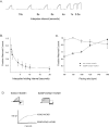
A) Response of S209P−KCNQ1+KCNE1 channels to depolarizations at various intervals. COS cells transfected with S209P−KCNQ1+KCNE1 were subjected to 1 second depolarizations to 80 mV with holding potentials of −80mV at the indicated durations. Figure shows one representative trace. B) The number of channels remaining open after holding potentials of different durations at −80mV. The height of the instantaneous step-up in current with depolarization at the end of the holding potential duration indicated on the X-axis was measured. Results were normalized to the maximal current obtained with a prolonged depolarization to 80mV for n=6 cells. C) Cells transfected with KCNQ1+KCNE1 or S209P− KCNQ1+KCNE1 were subjected to a 100 depolarizing pulses to +60mV at the indicated frequency. The amplitude of the current during the final depolarizing pulse was normalized to the maximal value for each channel. The effect of the voltage independent current carried by S209P−KCNQ1 channels is most marked at low stimulation frequencies. D) Deactivation kinetics of KCNQ1−KCNE1 and S209P KCNQ1−KCNE1 channels. The current traces shown are insets from the traces shown in figure 3, and show deactivation at −120mV. Kinetics for S209P KCNQ1−KCNE1 channels are significantly slower than wild-type channels.
Figure 6
Figure 6. Trafficking of S209P−KCNQ1 mutation for AF
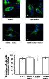
Confocal images of the KCNQ1 (A) and S209P (B) constructs in COS-7 cells. Membrane staining is noted. When the KCNQ1 (C) or S209P (D) constructs are co-expressed with KCNE1, there is again no difference in membrane staining between the wild-type and mutant constructs. The degree of membrane staining for each transfection is quantified in a blinded fashion as described in the methods section (E).

Comment in

  • Kv7.1 in atrial fibrillation.
    Seebohm G. Seebohm G. Heart Rhythm. 2009 Aug;6(8):1154-5. doi: 10.1016/j.hrthm.2009.05.004. Epub 2009 May 8. Heart Rhythm. 2009. PMID: 19560405 No abstract available.

References

    1. Go AS, Hylek EM, Phillips KA, et al. Prevalence of diagnosed atrial fibrillation in adults: national implications for rhythm management and stroke prevention: the AnTicoagulation and Risk Factors in Atrial Fibrillation (ATRIA) Study. JAMA. 2001;285(18):2370–2375. - PubMed
    1. Wang TJ, Larson MG, Levy D, et al. Temporal relations of atrial fibrillation and congestive heart failure and their joint influence on mortality: the Framingham Heart Study. Circulation. 2003;107(23):2920–2925. - PubMed
    1. Fox CS, Parise H, D'Agostino RB, Sr., et al. Parental atrial fibrillation as a risk factor for atrial fibrillation in offspring. JAMA. 2004;291(23):2851–2855. - PubMed
    1. Ellinor PT, Yoerger DM, Ruskin JN, MacRae CA. Familial aggregation in lone atrial fibrillation. Hum Genet. 2005;118(2):179–184. - PubMed
    1. Brugada R, Tapscott T, Czernuszewicz GZ, et al. Identification of a genetic locus for familial atrial fibrillation. N Engl J Med. 1997;336(13):905–911. - PubMed

Publication types