. 2009 Sep 18;284(38):25749-60.
doi: 10.1074/jbc.M109.042499. Epub 2009 Jul 24.

The Ca2+ affinity of synaptotagmin 1 is markedly increased by a specific interaction of its C2B domain with phosphatidylinositol 4,5-bisphosphate

Affiliations

The Ca2+ affinity of synaptotagmin 1 is markedly increased by a specific interaction of its C2B domain with phosphatidylinositol 4,5-bisphosphate

Anand Radhakrishnan et al. J Biol Chem. .

Abstract

The synaptic vesicle protein synaptotagmin 1 is thought to convey the calcium signal onto the core secretory machinery. Its cytosolic portion mainly consists of two C2 domains, which upon calcium binding are enabled to bind to acidic lipid bilayers. Despite major advances in recent years, it is still debated how synaptotagmin controls the process of neurotransmitter release. In particular, there is disagreement with respect to its calcium binding properties and lipid preferences. To investigate how the presence of membranes influences the calcium affinity of synaptotagmin, we have now measured these properties under equilibrium conditions using isothermal titration calorimetry and fluorescence resonance energy transfer. Our data demonstrate that the acidic phospholipid phosphatidylinositol 4,5-bisphosphate (PI(4,5)P2), but not phosphatidylserine, markedly increases the calcium sensitivity of synaptotagmin. PI(4,5)P2 binding is confined to the C2B domain but is not affected significantly by mutations of a lysine-rich patch. Together, our findings lend support to the view that synaptotagmin functions by binding in a trans configuration whereby the C2A domain binds to the synaptic vesicle and the C2B binds to the PI(4,5)P2-enriched plasma membrane.


Figures
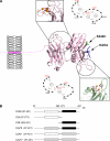
FIGURE 1.
FIGURE 1.
Structure of synaptotagmin 1. Synaptotagmin 1 protein consists of two C2 domains, C2A and C2B, that coordinate three and two calcium ions, respectively (16). The acidic residues that coordinate calcium binding is shown schematically, with the residues mutated in the calcium binding mutants (i.e. C2Ab*, C2a*B, and C2a*b*) shown in red. The Lys-rich patch is represented as a ball-and-stick model colored blue with the single cysteine site for the FRET assay (S342C) colored in green (A). The different mutants and constructs used in the study are schematically depicted (B).
FIGURE 2.
FIGURE 2.
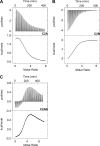
Calcium binding to the C2 domain of synaptotagmin 1 measured by ITC. Calcium chloride was titrated to 594 μm C2A domain (20 mm CaCl2) (A), 508 μm C2B domain (18 mm CaCl2) (B), and 500 μm wild-type C2AB (20 mm CaCl2) (C) at 25 °C in 50 mm HEPES, pH 7.4, 250 mm NaCl, 5 mm β-mercaptoethanol. The upper panels show the raw titration data, and the lower panels show the integrated heat changes after subtracting the heat of dilution. Interestingly, we observed that the two C2 domains of synaptotagmin adopt a thermodynamically divergent mechanism in calcium binding. The C2A domain bound calcium via an endothermic reaction, whereas the C2B domain exhibited an exothermic profile. The thermodynamic parameters of calcium binding are summarized in Table 1.
FIGURE 3.
FIGURE 3.
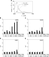
A novel FRET assay allows the monitoring of synaptotagmin 1 binding to liposomes. Binding was studied using FRET between synaptotagmin 1 labeled with the donor dye, Alexa 488, at position 342 on the C2B domain and liposomes containing phosphatidylethanolamine labeled with Texas Red as acceptor dye. Initially the spectrum was determined for the labeled synaptotagmin (0.2 μm) in the presence of 2 mm calcium (F0) (black (dotted line)). Upon the addition of liposomes (black (solid line)) and EGTA (gray), subsequent spectra are measured (F) (A). To compare the FRET changes for the different liposome samples, the fluorescence at 518 nm is normalized to the base0line value (F0/F). This normalization was done for the different synaptotagmin mutants with liposomes containing different compositions of lipids (i.e. 0, 10, and 25% phosphatidylserine in the absence (denoted as PS) or presence (denoted as PSP) of 1% PI(4,5)P2 (PIP2)) (B–E) (the color scheme is as in A). A.U., absorbance units. For all different liposomes tested, wild-type C2AB (B) exhibits a much stronger FRET signal than the calcium mutants C2a*B (C), C2Ab* (D), and C2a*b* (E). Note that the C2Ab* mutant appears to bind somewhat more efficiently to PI(4,5)P2-containing membranes but only at higher PS concentrations. It seems, therefore, possible that the mutated C2B domain of the C2Ab* mutant might still be able to contribute to membrane binding by interacting to some extent with PI(4,5)P2, hinting at a cooperative binding mechanism of calcium and PI(4,5)P2. The C2a*b* variant, which does not bind calcium (supplemental Fig. 1), did not exhibit any detectable binding to the different liposomes.
FIGURE 4.
FIGURE 4.
The two C2 domains of synaptotagmin 1 cooperate for membrane binding. Texas Red-labeled liposomes containing 25% phosphatidylserine were titrated to 0.2 μm Alexa 488-labeled synaptotagmin in the presence of saturating amounts of calcium (2 mm). Titrations were done with liposomes with (○) and without (●) 1% PI(4,5)P2 for the wild-type C2AB (A) and the calcium mutants C2a*B (B) and C2Ab* (C). When the titration was carried out the presence of 50 μm calcium chloride, hardly any binding of synaptotagmin to liposomes without PI(4,5)P2 was observed (D). The PI(4,5)P2-containing liposomes on the other hand are able to still bind synaptotagmin, albeit with reduced affinity (EC50 = 7.0 μm), compared with saturating calcium concentrations (2 mm; EC50 = 2.4 μm). The fits to the Hill function are shown by continuous lines. It is possible that the higher FRET signal observed for the intact protein compared with the Ca2+ mutants in part arises from its deeper penetration into the membrane (as reported by Herrick et al. (21) for the C2AB protein), although this is difficult to confirm in our present study. For each titration the relative fluorescence was plotted against the PS concentration. The PS concentration was calculated from the total lipid concentration, which was determined by measuring the total phosphate content of the liposome sample. Notably, the global membrane binding affinity determined for synaptotagmin is in a similar range found for classical PKC C2 domains (26, 27).
FIGURE 5.
FIGURE 5.
Calcium affinity of synaptotagmin is augmented by PI(4,5)P2. An excess (∼0.4 mm) of Texas Red-labeled liposomes was mixed with 0.2 μm Alexa 488-labeled synaptotagmin in 50 mm HEPES, pH 7.4, 150 mm NaCl containing 10 mm DPTA (KD = 80 μm) as a chelator to ensure accurate free calcium concentrations. Calcium was then added stepwise, and the fluorescence quenching of the donor dye that denotes binding of synaptotagmin to membranes was recorded. The free calcium concentrations were calculated using the Igor Pro software and plotted against the relative fluorescence change. Calcium titrations were done for the wild-type C2AB (A) and the calcium mutants C2a*B (B) and C2Ab* (C) using liposomes with (○) and without (●) 1% PI(4,5)P2.
FIGURE 6.
FIGURE 6.
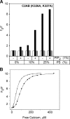
The KAKA mutant behaves similar to the wild-type synaptotagmin protein. Membrane binding of wild-type or KAKA mutant (K326A, K327A) of synaptotagmin 1 was compared using the FRET assay described in Figs. 3 and 5 (A). Liposomes with different lipid compositions were tested (B) (with similar schemes as described for Fig. 3). For calcium titrations, liposomes with (○) and without (●) 1% PI(4,5)P2 (PIP2) were used.
FIGURE 7.
FIGURE 7.
Effects of Ca2+ on the fusion of synaptobrevin liposomes containing full-length synaptotagmin with Q-SNARE liposomes in the presence or absence of PI(4,5)P2. Membrane-inserted synaptotagmin is able to stimulate the process of SNARE protein mediated membrane fusion in the presence of Ca2+ when only the Q-SNARE liposome membrane contains the acidic lipid phosphatidylserine (24). To test for the effect of Ca2+ on the fusion process, the Q-SNARE liposome membrane contained 10% PS in the absence or presence of 1% PI(4,5)P2, whereas the liposomes containing synaptobrevin did not contain PS. Fusion between syntaxin 1a-SNAP-25-containing liposomes and synaptobrevin-containing liposomes was measured by a standard lipid dequenching assay. Fluorescence values were normalized to the initial fluorescence measured (denoted as F/F0). Individual fusion reactions were carried out at different calcium concentrations and repeated three times, each time using freshly prepared liposomes. Selected kinetic traces are shown in supplemental Fig. 4. Although the kinetics were rather complex, i.e. composed of at least two different phases, in the presence of 1% PI(4,5)P2 much less Ca2+ is needed to increase the efficiency of membrane fusion. To evaluate the stimulating effect, the amount of fusion in each reaction was plotted after 300 s.

Similar articles

Cited by

References

    1. Sudhof T. C. (2004) Annu. Rev. Neurosci. 27, 509–547 - PubMed
    1. Jahn R., Scheller R. H. (2006) Nat. Rev. Mol. Cell Biol. 7, 631–643 - PubMed
    1. Rizo J., Chen X., Araç D. (2006) Trends Cell Biol. 16, 339–350 - PubMed
    1. Wojcik S. M., Brose N. (2007) Neuron 55, 11–24 - PubMed
    1. Chapman E. R. (2008) Annu. Rev. Biochem. 77, 615–641 - PubMed

MeSH terms