

Use of uteroglobin for the engineering of polyvalent, polyspecific fusion proteins
- PMID: 19632988
- PMCID: PMC2785352
- DOI: 10.1074/jbc.M109.025924
Use of uteroglobin for the engineering of polyvalent, polyspecific fusion proteins
Abstract
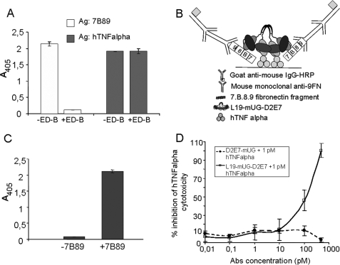
We report a novel strategy to engineer and express stable and soluble human recombinant polyvalent/polyspecific fusion proteins. The procedure is based on the use of a central skeleton of uteroglobin, a small and very soluble covalently linked homodimeric protein that is very resistant to proteolytic enzymes and to pH variations. Using a human recombinant antibody (scFv) specific for the angiogenesis marker domain B of fibronectin, interleukin 2, and an scFv able to neutralize tumor necrosis factor-alpha, we expressed various biologically active uteroglobin fusion proteins. The results demonstrate the possibility to generate monospecific divalent and tetravalent antibodies, immunocytokines, and dual specificity tetravalent antibodies. Furthermore, compared with similar fusion proteins in which uteroglobin was not used, the use of uteroglobin improved properties of solubility and stability. Indeed, in the reported cases it was possible to vacuum dry and reconstitute the proteins without any aggregation or loss in protein and biological activity.
Figures


Similar articles
-
Selective targeted delivery of the TNF-alpha receptor p75 and uteroglobin to the vasculature of inflamed tissues: a preliminary report.BMC Biotechnol. 2011 Nov 10;11:104. doi: 10.1186/1472-6750-11-104. BMC Biotechnol. 2011. PMID: 22074550 Free PMC article.
-
Use of the uteroglobin platform for the expression of a bivalent antibody against oncofetal fibronectin in Escherichia coli.PLoS One. 2013 Dec 19;8(12):e82878. doi: 10.1371/journal.pone.0082878. eCollection 2013. PLoS One. 2013. PMID: 24367567 Free PMC article.
-
A new recombinant single chain trispecific antibody recruits T lymphocytes to kill CEA (carcinoma embryonic antigen) positive tumor cells in vitro efficiently.J Biochem. 2004 Apr;135(4):555-65. doi: 10.1093/jb/mvh065. J Biochem. 2004. PMID: 15115782
-
Pharmacokinetics and biodistribution of genetically-engineered antibodies.Q J Nucl Med. 1998 Dec;42(4):225-41. Q J Nucl Med. 1998. PMID: 9973838 Review.
-
Generation of humanized monoclonal antibodies by 'best fit' framework selection and recombinant polymerase chain reaction.Year Immunol. 1993;7:110-8. Year Immunol. 1993. PMID: 8372500 Review. No abstract available.
Cited by
-
Selective targeted delivery of the TNF-alpha receptor p75 and uteroglobin to the vasculature of inflamed tissues: a preliminary report.BMC Biotechnol. 2011 Nov 10;11:104. doi: 10.1186/1472-6750-11-104. BMC Biotechnol. 2011. PMID: 22074550 Free PMC article.
-
Use of the uteroglobin platform for the expression of a bivalent antibody against oncofetal fibronectin in Escherichia coli.PLoS One. 2013 Dec 19;8(12):e82878. doi: 10.1371/journal.pone.0082878. eCollection 2013. PLoS One. 2013. PMID: 24367567 Free PMC article.
-
Antibody vectors for imaging.Semin Nucl Med. 2010 May;40(3):167-81. doi: 10.1053/j.semnuclmed.2009.12.005. Semin Nucl Med. 2010. PMID: 20350626 Free PMC article. Review.
References
-
- Krishnan R. S., Daniel J. C., Jr. (1967) Science 158, 490–492 - PubMed
-
- Beier H. M. (1968) Biochim. Biophys. Acta 160, 289–291 - PubMed
-
- Klug J., Beier H. M., Bernard A., Chilton B. S., Fleming T. P., Lehrer R. I., Miele L., Pattabiraman N., Singh G. (2000) Ann. N.Y. Acad. Sci. 923, 348–354 - PubMed
-
- Morize I., Surcouf E., Vaney M. C., Epelboin Y., Buehner M., Fridlansky F., Milgrom E., Mornon J. P. (1987) J. Mol. Biol. 194, 725–739 - PubMed
-
- Mukherjee A. B., Chilton B. S. (2000) The Uterogloglobin/Clara Cell Protein Family, The New York Academy of Sciences, New York
Publication types
MeSH terms
Substances
LinkOut - more resources
Full Text Sources
Other Literature Sources
