. 2009 Sep 18;284(38):25782-90.
doi: 10.1074/jbc.M109.017194. Epub 2009 Jul 24.

Dual acylation of PDE2A splice variant 3: targeting to synaptic membranes

Affiliations

Dual acylation of PDE2A splice variant 3: targeting to synaptic membranes

Corina Russwurm et al. J Biol Chem. .

Abstract

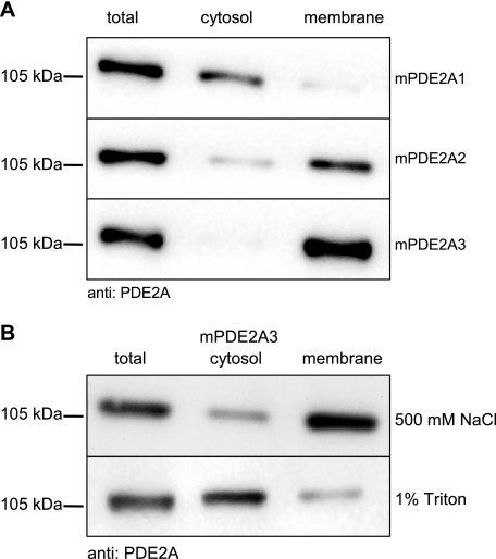
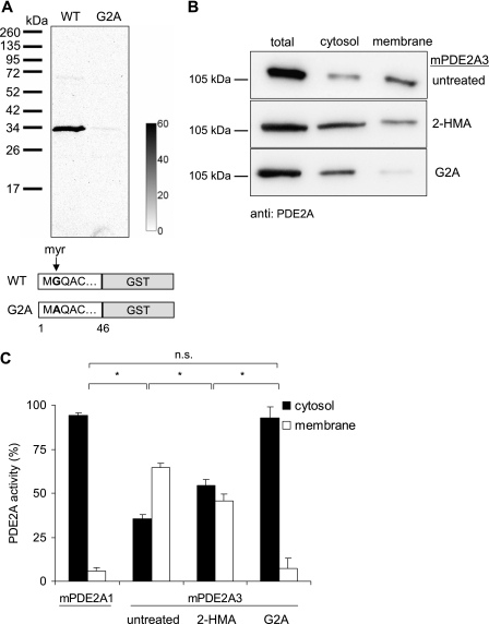
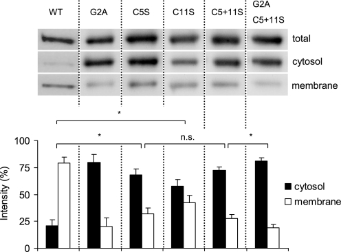
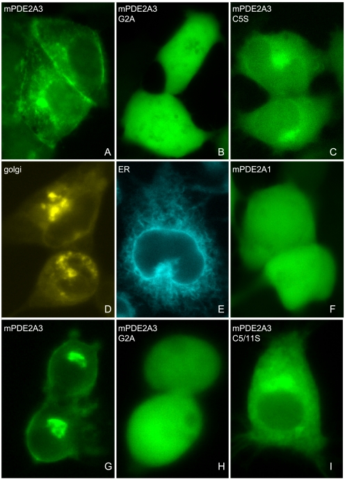
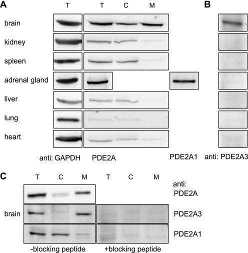
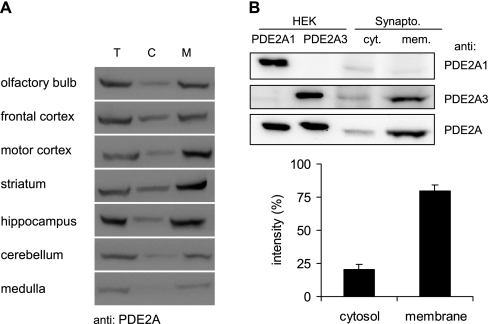
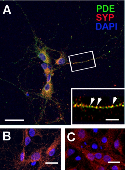
The cGMP-stimulated PDE2A hydrolyzes both cyclic nucleotides, cGMP and cAMP. Three splice variants have been cloned from several species. Whereas PDE2A1 is soluble, PDE2A2 and PDE2A3 are membrane-bound enzymes of rat and bovine origin, respectively. To date it is unclear whether one species expresses all three variants. The splice variants only differ in their N termini, which likely determine the subcellular localization. However, the mechanism for membrane attachment remains unknown. Here, we show that myristoylation underlies membrane targeting of PDE2A3. The myristoylated enzyme was bound to plasma membranes, whereas mutation of the myristoyl recipient Gly2 prevented incorporation of [3H]myristate and turned PDE2A3 completely soluble. Additionally, Cys5 and to a minor extent Cys11 are required for targeting of PDE2A3. Substitution of the putatively palmitoylated cysteines partially solubilized the enzyme and led to an accumulation in the endoplasmic reticulum/Golgi compartment, as shown by fluorescence microscopy in HEK 293 and PC12 cells. In vivo, PDE2A is expressed in many tissues. By using newly generated antibodies selectively detecting the splice variants PDE2A3 or PDE2A1, respectively, we demonstrate on the protein level PDE2A3 expression in mouse brain where it is entirely membrane-associated and a widespread expression of soluble PDE2A1 in mouse tissues. We show that PDE2A localizes to synaptosomal membranes and in primary cultures of hippocampal neurons partially overlaps with the presynaptic marker synaptophysin as demonstrated by immunofluorescence. In sum, these results demonstrate dual acylation as mechanism targeting neuronal PDE2A3 to synapses thereby ensuring local control of cyclic nucleotides.


Figures

FIGURE 1.
FIGURE 1.
Subcellular localization of PDE2A splice variants. A, HEK 293 cells were transiently transfected with the PDE2A splice variants. Cells were homogenized and separated into cytosolic and membrane fractions as described under “Materials and Methods.” Western blot analysis was performed using anti-PDE2A antibody. B, PDE2A3-expressing HEK 293 cells were homogenized in the presence of 500 mm NaCl (top) or 1% Triton X-100 (bottom). Cytosolic and membrane fractions were analyzed in Western blots. Shown are representative results of three independent experiments.
FIGURE 2.
FIGURE 2.
N-Myristoylation of PDE2A3. A, GST fusion proteins of the N-terminal 46 amino acids of PDE2A3 or its G2A mutant, respectively, were labeled with [3H]myristic acid in HEK 293 cells. After precipitation, electrophoresis, and blotting, labeled proteins were detected using a phosphorimaging device. The calibration bar depicts mPSL units (photostimulated luminescence units, Fujifilm). B, Western blot of cytosolic and membrane fractions of PDE2A3-expressing HEK 293 cells. Top, untreated cells. Middle, cells treated with the myristoylation inhibitor 2-HMA (0.5 mm). Bottom, untreated cells expressing the myristoylation deficient G2A mutant of PDE2A3. C, PDE activities in subcellular fractions of HEK 293 cells expressing PDE2A1, PDE2A3, or the G2A mutant of PDE2A3. PDE2A3-expressing cells were treated with 0.5 mm 2-HMA as indicated. Shown are means ± S.D. of at least three independent experiments performed in duplicate; n.s., not significant (p > 0.05); *, significant (p < 0.003), two-tailed t test assuming unequal variances.
FIGURE 3.
FIGURE 3.
Cysteines-5 and -11 contribute to membrane attachment of PDE2A3. PDE2A3 carrying point mutations at positions 2 (G2A), 5 (C5S), and 11 (C11S) or a combination thereof, were transiently transfected into HEK 293 cells. Equal volumes of separated cell fractions were then blotted and probed with the PDE2A-specific antibody. Shown is a representative blot of three independent experiments. For comparison, the PDE2A3 WT is shown on the left. The lower panel shows the quantitative analysis of PDE2A3 signal intensities in cytosol and membrane fractions, which together were set to 100%. Bars are means ± S.D. of three independent experiments; n.s., not significant (p > 0.05); *, significant (p < 0.01), two-tailed paired t test.
FIGURE 4.
FIGURE 4.
Membrane targeting of a PDE2A3/CFP fusion protein is disrupted by G2A and C5S mutations in HEK 293 and PC12 cells. Cells were grown on coverslips and transfected with the PDE2A3 N terminus fused to CFP. A–F, expression in HEK 293 cells. G–I, distribution of the denoted CFP fusion proteins in PC12 cells. A, wild-type PDE2A3 N terminus was fused to CFP. B, Gly2 of the PDE2A3 N terminus was changed to Ala. C, Cys5 of the PDE2A3 N terminus was changed to Ser. D, Golgi marker vector pECFP-Golgi (Clontech). E, ER marker pECFP-ER (Clontech). F, as an example of a soluble enzyme a PDE2A1/CFP fusion protein was expressed.
FIGURE 5.
FIGURE 5.
Tissue distribution of PDE2A splice variants in mouse. A, mouse organs were processed by means of a glass-Teflon homogenizer and fractionated by centrifugation. PDE2A localization was then analyzed by SDS-PAGE and Western blotting using the PDE2A-specific antibody. Due to the very limited amount of material, adrenal gland homogenate was not fractionated but probed with isoform-specific antibodies detecting PDE2A1 or PDE2A3 (see B). For an internal standard glyceraldehyde-3-phosphate dehydrogenase (GAPDH) was detected in the homogenates. B, homogenates of mouse organs were probed using an antibody specifically detecting PDE2A3. C, mouse brain was processed as described above using buffer containing 150 mm NaCl. 25 μg of total protein was blotted, and PDE2A was detected using antibodies as indicated. On the right, isoform-specific detection in the presence of the respective blocking peptide is shown. T, total; C, cytosol; and M, membrane.
FIGURE 6.
FIGURE 6.
Neuronal expression of PDE2A. A, the denoted areas of mouse brains were separated into soluble and particulate fractions. 20 μg of total protein per lane were separated by SDS-PAGE. For Western detection, the PDE2A-specific antibody was used. T, total; C, cytosol; M, membrane. B, crude synaptosomal vesicles obtained from mouse brains were separated into cytosol and membranes and subjected to Western blots using isoform-specific antibodies against PDE2A1 and PDE2A3 and a pan-specific antibody (PDE2A) recognizing both isoforms. For comparison, homogenates of HEK cells expressing either PDE2A1 or PDE2A3 are shown. Quantitative analysis of pan-specific PDE2A signals from three independent experiments is displayed graphically with the signal intensity of the cytosol plus membrane set to 100%. Bars are means ± S.D. of three independent experiments; p < 0.01 (two-tailed paired t test).
FIGURE 7.
FIGURE 7.
PDE2A and synaptophysin co-localization in primary hippocampal neurons. A, rat hippocampal neurons isolated as described under “Materials and Methods” and fixed for immunohistochemistry were double labeled with PDE2A (PDE, green), synaptophysin (SYP, red), and Alexa Fluor-conjugated secondary antibodies. The nuclei were stained with 4′,6-diamidino-2-phenylindole (DAPI). Neurons were examined by confocal microscopy. The inset demonstrates the distribution of PDE2A and SYP within a single neuronal process and indicates co-localizing puncta (arrowheads). B, the PDE2A-specific signal was blocked by incubation with blocking peptide. C, control with secondary antibody alone. Scale bars represent 30 μm (A), 6 μm (inset), and 20 μm (B and C).

Similar articles

Cited by

References

    1. Bender A. T., Beavo J. A. (2006) Pharmacol. Rev. 58, 488–520 - PubMed
    1. Martins T. J., Mumby M. C., Beavo J. A. (1982) J. Biol. Chem. 257, 1973–1979 - PubMed
    1. Beavo J. A., Hardman J. G., Sutherland E. W. (1971) J. Biol. Chem. 246, 3841–3846 - PubMed
    1. Erneux C., Couchie D., Dumont J. E., Baraniak J., Stec W. J., Abbad E. G., Petridis G., Jastorff B. (1981) Eur. J. Biochem. 115, 503–510 - PubMed
    1. MacFarland R. T., Zelus B. D., Beavo J. A. (1991) J. Biol. Chem. 266, 136–142 - PubMed

Publication types

MeSH terms