. 2009 Oct 2;284(40):27243-51.
doi: 10.1074/jbc.M109.032565. Epub 2009 Jul 23.

Mapping the cargo protein membrane translocation step into the PEX5 cycling pathway

Affiliations

Mapping the cargo protein membrane translocation step into the PEX5 cycling pathway

Inês S Alencastre et al. J Biol Chem. .

Abstract

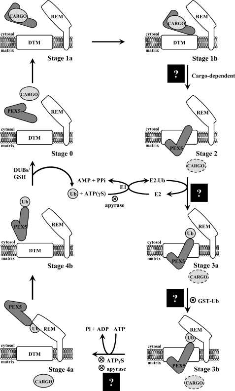
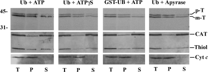
Newly synthesized peroxisomal matrix proteins are targeted to the organelle by PEX5, the peroxisomal cycling receptor. Over the last few years, valuable data on the mechanism of this process have been obtained using a PEX5-centered in vitro system. The data gathered until now suggest that cytosolic PEX5.cargo protein complexes dock at the peroxisomal docking/translocation machinery, where PEX5 becomes subsequently inserted in an ATP-independent manner. This PEX5 species is then monoubiquitinated at a conserved cysteine residue, a mandatory modification for the next step of the pathway, the ATP-dependent dislocation of the ubiquitin-PEX5 conjugate back into the cytosol. Finally, the ubiquitin moiety is removed, yielding free PEX5. Despite its usefulness, there are many unsolved mechanistic aspects that cannot be addressed with this in vitro system and that call for a cargo protein-centered perspective instead. Here we describe a robust peroxisomal in vitro import system that provides this perspective. The data obtained with it suggest that translocation of a cargo protein across the peroxisomal membrane, including its release into the organelle matrix, occurs prior to PEX5 ubiquitination.


Figures

FIGURE 1.
FIGURE 1.
The PEX5-mediated peroxisomal protein import pathway. There are five major stages in this protein sorting pathway (numbered 0–4). Substages (a and b) are mostly of conceptual nature. The different stages have been characterized with a PEX5-centered in vitro system, applying several strategies that block (⊗) the pathway at different steps. Stage 0, cytosolic cargo-free PEX5. Stage 1, cytosolic PEX5·cargo protein complex. Stage 2, PEX5 embedded in the peroxisomal DTM. Stage 3, DTM-embedded monoubiquitinated PEX5. Stage 4, cytosolic monoubiquitinated PEX5. Insertion of PEX5 into the DTM is cargo protein-dependent. Monoubiquitination of stage 2 PEX5 yielding stage 3 PEX5 requires activated ubiquitin. Activation of ubiquitin by the ubiquitin-activating enzyme (E1) is an ATP-dependent process; E2 indicates ubiquitin carrier protein. Note that this activation, which involves the synthesis of the acyl phosphate AMP-ubiquitin anhydride and the release of pyrophosphate, can also be achieved with ATPγS (a sulfur atom at a non-bridge position of the γ-phosphate of ATP does not affect the reaction (62)). The ubiquitin analogue GST·Ub is also used efficiently by the ubiquitin-conjugating cascade acting on PEX5. However, this stage 3 species is no longer a substrate for the receptor export module (REM), presumably because of the bulkiness of GST·Ub. Apyrase hydrolyzes ATP and thus blocks PEX5 both at stage 2 and stage 3b levels. Note that if apyrase is added to the in vitro assays before PEX5 (or ΔC1PEX5L), the receptor will not proceed to stage 3a. Stage 4 PEX5 is deubiquitinated, yielding stage 0 PEX5 in a process that probably involves deubiquitinating enzymes (DUBs) and GSH. The step at which cargo proteins are translocated across the peroxisomal membrane (black squares with a question mark) is the topic of this work. Mammalian PEX7 is not represented in the model because it is presently unknown whether it is retained at the DTM together with PEX5L or translocated across the peroxisomal membrane together with the cargo protein, as proposed for the yeast protein (63).
FIGURE 2.
FIGURE 2.
A robust amount of 35S-labeled prethiolase acquires a protease-protected and organelle-associated status in in vitro import reactions fortified with ΔC1PEX5L or PEX5L(N526K). A, a rat liver PNS fraction was incubated with 35S-labeled prethiolase in import buffer containing ATP in the absence (lane 2) or presence of recombinant ΔC1PEX5L (lane 3), ΔC1PEX5S (lane 4), PEX5L (lane 5), or PEX5L(N526K) (lane 6). After trypsin treatment, the organelles were isolated by centrifugation, subjected to SDS-PAGE, and blotted onto a nitrocellulose membrane. The Ponceau S-stained membrane (lower panel) and its autoradiograph (upper panel) are shown. Lane 1, 10% of the reticulocyte lysate containing 35S-labeled prethiolase used in each reaction. B, time dependence of the ΔC1PEX5L-mediated 35S-labeled prethiolase import. Aliquots from a 5-fold standard import reaction were withdrawn at the indicated time points and processed as described in A. The Ponceau S-stained membrane (lower panel) and its autoradiograph (upper panel) are shown. Lane 1, 10% of the reticulocyte lysate containing 35S-labeled prethiolase used in each lane. C, import of 35S-labeled prethiolase is temperature-dependent. A PNS pretreated with 0.3 mm ATP for 5 min at 37 °C (see “Experimental Procedures”) was used in import reactions containing ATP and recombinant ΔC1PEX5L. After 45 min at the indicated temperatures, the samples were processed as described in A. The Ponceau S-stained membrane (lower panel) and its autoradiograph (upper panel) are shown. Lane 1, 10% of the reticulocyte lysate containing 35S-labeled prethiolase used in each reaction. D, 35S-labeled thiolase lacking its PTS2 does not acquire a protease-resistant/organelle-associated status when incubated with ΔC1PEX5L-supplemented rat liver PNS. Standard import reactions supplemented with ΔC1PEX5L (lanes 4 and 6) or lacking this recombinant protein (lanes 3 and 5) were programmed with 35S-labeled prethiolase (lanes 3 and 4) or 35S-labeled thiolase lacking the PTS2 (ΔPTS2-T; lanes 5 and 6). At the end of the incubation, the samples were processed as described in A. The Ponceau S-stained membrane (lower panel) and its autoradiograph (upper panel) are shown. Lane 1, 35S-labeled prethiolase (10% of the input used in lanes 3 and 4); lane 2, 35S-labeled thiolase lacking the PTS2 (10% of the input used in lanes 5 and 6). p-T and m-T, precursor and mature forms of thiolase, respectively. The asterisk marks a radiolabeled band produced by the in vitro translation kit in an unspecific manner. The numbers at the left indicate the molecular masses of the applied standards in kDa.
FIGURE 3.
FIGURE 3.
35S-Labeled prethiolase is specifically imported into peroxisomes. A ΔC1PEX5L-supplemented PNS fraction was incubated with 35S-labeled prethiolase in import buffer containing ATP for 45 min. After trypsin treatment and inactivation of the protease, the complete import mixture was diluted with SEM buffer and subjected to Nycodenz gradient centrifugation. The gradient was then fractionated from the bottom (lane 1) to the top (lane 14), and equal aliquots from each fraction were subjected to SDS-PAGE and Western blotting. The nitrocellulose membrane was exposed to an x-ray film to detect the 35S-labeled protein (top panel) and afterward probed with the following antisera: anti-KDEL (KDEL; recognizes GRP72 and GRP98, two endoplasmic reticulum proteins), anti-cytochrome c (Cyt c; a mitochondrial marker), anti-catalase (CAT; a peroxisomal enzyme), and anti-PEX13. This last serum recognizes on trypsin-treated peroxisomes a 30-kDa fragment of PEX13 (PEX13′) (46). Note that catalase remaining at the top of the gradient (lanes 11–14) results from leakage of peroxisomes during preparation of PNS fractions. p-T and m-T, precursor and mature forms of thiolase, respectively. The numbers at the right indicate the molecular masses of the applied standards in kDa.
FIGURE 4.
FIGURE 4.
Import of prethiolase into peroxisomes does not require hydrolysis of cytosolic ATP. A, import reactions assembled with components pretreated with 0.3 mm ATP (see “Experimental Procedures”) and supplemented with either bovine Ub or GST·Ub were performed in the presence of 3 mm ATP or 3 mm ATPγS, as indicated. After trypsin treatment, the organelles were isolated by centrifugation and analyzed as described in the legend for Fig. 2. The autoradiograph (upper panel) and the corresponding Ponceau S-stained membrane (lower panel) are shown. Lane 1, 10% of 35S-labeled prethiolase solution used in each lane. B, a rat liver PNS was pretreated with 0.3 mm ATP for 5 min and divided into four equal aliquots (lanes 2–5). The first and second aliquot (lanes 2 and 3, respectively) received bovine ubiquitin and 3 mm ATP; the third aliquot received GST·Ub and 3 mm ATP (lane 4); and the fourth received GST·Ub and apyrase (Apy). After 3 min at 37 °C, the first aliquot received 35S-labeled prethiolase preincubated with ATP in the absence of ΔC1PEX5L, whereas the second and third aliquots received 35S-labeled prethiolase preincubated with ATP in the presence of ΔC1PEX5L (lanes 3 and 4). The fourth aliquot received 35S-labeled prethiolase preincubated with ATP plus ΔC1PEX5L and treated with apyrase (see “Experimental Procedures” for details). After 45 min at 37 °C, the samples were treated with trypsin and processed as described above. Lane 1, 10% of the 35S-labeled prethiolase used in the import reactions. The autoradiograph (upper panel) and the corresponding Ponceau S-stained membrane (lower panel) are shown. p-T and m-T, precursor and mature forms of thiolase, respectively. C, in vitro assays using 35S-labeled PEX5L(C11K). The experimental conditions used in lanes 2 and 3 were exactly the ones used in lanes 4 and 5 of the experiment shown in B, respectively, with the exception that recombinant ΔC1PEX5L was omitted because it strongly competes with the radiolabeled protein for the DTM. At the end of the 45-min incubation at 37 °C, the organelles were sedimented and processed for SDS-PAGE. Note that PEX5L(C11K) is as functional as normal PEX5L in these assays (23). It was used here for practical reasons because the GST·Ub·PEX5L(C11K) conjugate, unlike the GST·Ub·PEX5L, is not destroyed by prolonged incubation in the presence of GSH and can be analyzed under normal SDS-PAGE conditions (23). The complete absence of GST·Ub·PEX5L(C11K) in lane 3 indicates that the amount of apyrase used in these assays efficiently depletes ATP from the reactions. Lane 1, 10% of 35S-labeled PEX5L(C11K) used in each reaction. Numbers to the left indicate the molecular masses of protein standards in kDa.
FIGURE 5.
FIGURE 5.
Release of in vitro imported 35S-labeled thiolase into the peroxisomal matrix occurs before stage 3. Trypsin-treated organelles from in vitro import reactions, containing the indicated combinations of bovine Ub, GST·Ub, ATP, ATPγS, or apyrase and performed as described in legend for Fig. 4, were disrupted by sonication in a low ionic strength buffer and divided into two halves. One-half (samples T) was kept on ice, whereas the other was subjected to ultracentrifugation to separate membranes (samples P) from soluble proteins (samples S). Equivalent portions of samples T, P, and S were subjected to SDS-PAGE and blotted onto a nitrocellulose. The membrane was first exposed to an x-ray film to detect the 35S-labeled protein (top panel) and afterward probed with the following antisera: anti-thiolase (Thiol); anti-catalase (CAT), and anti-cytochrome c (Cyt c). p-T and m-T, precursor and mature forms of thiolase, respectively. Numbers to the left indicate the molecular masses of protein standards in kDa.

Similar articles

Cited by

References

    1. Purdue P. E., Lazarow P. B. (2001) Annu. Rev. Cell Dev. Biol. 17, 701–752 - PubMed
    1. Brown L. A., Baker A. (2008) Mol. Membr. Biol. 25, 363–375 - PubMed
    1. Gould S. J., Keller G. A., Hosken N., Wilkinson J., Subramani S. (1989) J. Cell Biol. 108, 1657–1664 - PMC - PubMed
    1. Brocard C., Hartig A. (2006) Biochim. Biophys. Acta 1763, 1565–1573 - PubMed
    1. Swinkels B. W., Gould S. J., Bodnar A. G., Rachubinski R. A., Subramani S. (1991) EMBO J. 10, 3255–3262 - PMC - PubMed

Publication types

Substances

LinkOut - more resources