. 2009 Sep 18;284(38):26194-206.
doi: 10.1074/jbc.M109.019836. Epub 2009 Jul 24.

Silencing cAMP-response element-binding protein (CREB) identifies CYR61 as a tumor suppressor gene in melanoma

Affiliations

Silencing cAMP-response element-binding protein (CREB) identifies CYR61 as a tumor suppressor gene in melanoma

Andrey S Dobroff et al. J Biol Chem. .

Abstract

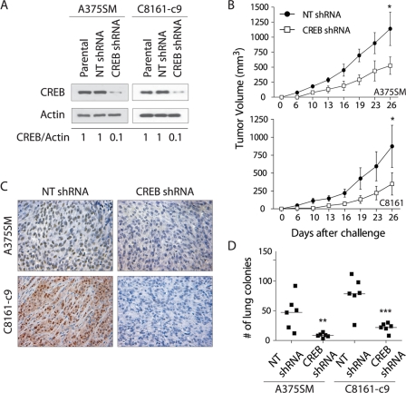
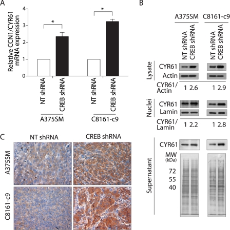
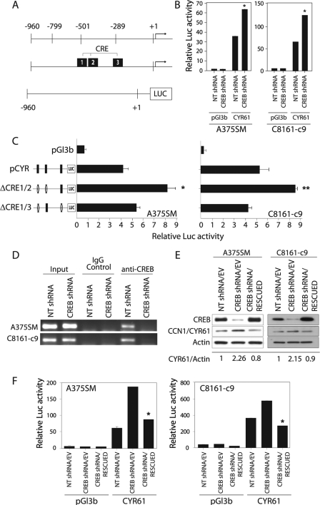
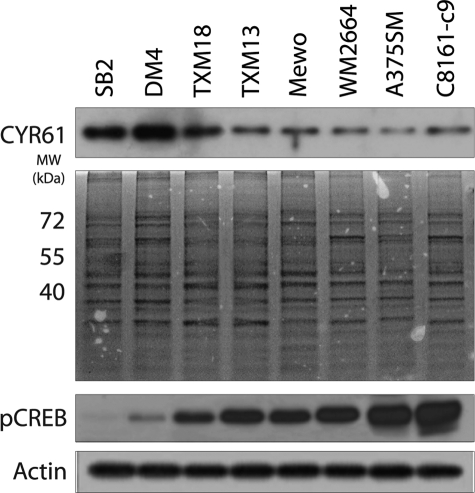
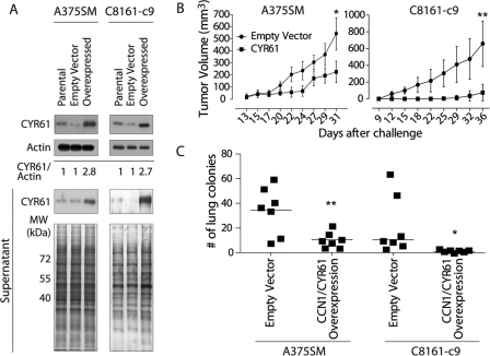
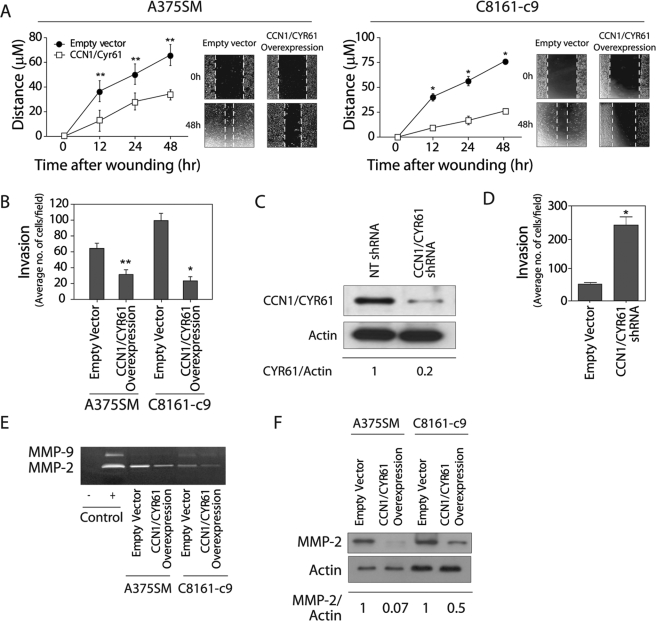
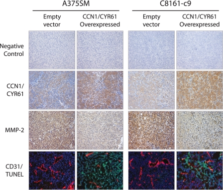
Metastatic progression of melanoma is associated with overexpression and activity of cAMP-response element-binding protein (CREB). However, the mechanism by which CREB contributes to tumor progression and metastasis remains unclear. Here, we demonstrate that stably silencing CREB expression in two human metastatic melanoma cell lines, A375SM and C8161-c9, suppresses tumor growth and experimental metastasis. Analysis of cDNA microarrays revealed that CREB silencing leads to increased expression of cysteine-rich protein 61 (CCN1/CYR61) known to mediate adhesion, chemostasis, survival, and angiogenesis. Promoter analysis and chromatin immunoprecipitation assays demonstrated that CREB acts as a negative regulator of CCN1/CYR61 transcription by directly binding to its promoter. Re-expression of CREB in CREB-silenced cells rescued the low CCN1/CYR61 expression phenotype. CCN1/CYR61 overexpression resulted in reduced tumor growth and metastasis and inhibited the activity of matrix metalloproteinase-2. Furthermore, its overexpression decreased melanoma cell motility and invasion through Matrigel, which was abrogated by silencing CCN1/CYR61 in low metastatic melanoma cells. Moreover, a significant decrease in angiogenesis as well as an increase in apoptosis was seen in tumors overexpressing CCN1/CYR61. Our results demonstrate that CREB promotes melanoma growth and metastasis by down-regulating CCN1/CYR61 expression, which acts as a suppressor of melanoma cell motility, invasion and angiogenesis.


Figures

FIGURE 1.
FIGURE 1.
Effects of CREB silencing on melanoma growth and metastasis. A, Western blot analysis shows silencing of CREB in the A375SM and C8161-c9 cell lines stably transduced with CREB-shRNA as compared with those transduced with nontargeting control shRNA (NT shRNA). α-Actin was used as a loading control. A 90% reduction in CREB was observed in the CREB-shRNA-transduced cells as compared with the NT-shRNA-transduced cells as measured by densitometry (0.1 and 1, respectively). B, silencing of CREB resulted in a significant inhibition of tumor growth in both the A375SM (CREB-shRNA, 278.1 mm3; NT-shRNA, 1140.0 mm3) and C8161-c9 (CREB-shRNA, 348.4 mm3; NT-shRNA, 873.0 mm3) melanoma cell lines. *, p < 0.05 for both A375SM and C8161-c9 cells. C, immunohistochemical staining for CREB was performed on tumor samples from mice 26 days after injection with CREB-shRNA- or NT-shRNA-transduced A375SM or C8161-c9 cells, demonstrating down-regulation of CREB expression in CREB-shRNA tumors. Images are shown at ×20 magnification. D, effect of CREB silencing on the metastatic potential of cells. There is a significant decrease in the number of lung metastases in CREB-shRNA-transduced A375SM and C8161-c9 cells as compared with the NT-shRNA-transduced cells. Each square represents one mouse (n = 6/group). **, p < 0.01; ***, p < 0.001.
FIGURE 2.
FIGURE 2.
Validation of CCN1/CYR61 overexpression after CREB silencing in melanoma cells. A, quantitative real-time PCR validation for CCN1/CYR61 gene expression. Expression values shown are -fold change in each CREB-shRNA-transduced cell line relative to the NT-shRNA cells after normalization with 18s RNA. *, p < 0.005. B, Western blot analyses of total cell lysate, nuclei and supernatant show a significant increase in CCN1/CYR61 expression in both A375SM and C8161-c9 cell lines after CREB silencing as compared with NT-shRNA cells. α-Actin, lamin, and whole gel staining were used as indicators of equal sample loading. C, immunohistochemical staining for CCN1/CYR61 was performed on tumor samples from mice 26 days after injection with NT-shRNA- or CREB-shRNA-transduced A375SM or C8161-c9 cells, demonstrating CCN1/CYR61 overexpression in CREB-shRNA-transduced cells in vivo.
FIGURE 3.
FIGURE 3.
Regulation of the CCN1/CYR61 promoter by CREB. A, schematic representation of the CCN1/CYR61 promoter region fused to the luciferase reporter gene and its predicted CRE binding sites. B, the luciferase activity driven by the CCN1/CYR61 promoter increased 2-fold after CREB silencing in both the A375SM and C8161-c9 cell lines as compared with the NT-shRNA-transduced cells. *, p < 0.001. C, a schematic representation of the promoter point mutations is depicted on the left side of the panel. The three identified CRE binding sites were mutated either alone or in combination, as described under “Experimental Procedures.” The luciferase activity driven by the CCN1/CYR61 promoter increased 2.2-fold when both the first and second CRE binding sites were mutated in the A375SM and C8161-c9 cell lines. *, p < 0.001; ** p < 0.01. D, chromatin immunoprecipitation studies showed no binding of CREB to the CCN1/CYR61 promoter in either of the CREB-silenced cell lines (A375SM and C8161-c9). IgG antibodies were used as negative controls. Input DNA was used to ensure an equal amount of chromatin used in each assay. E, rescue of CREB expression in the CREB-silenced cells results in down-regulation of CCN1/CYR61 expression. α-Actin was used as a loading control. F, the luciferase activity driven by the CCN1/CYR61 promoter decreased significantly (*, p < 0.001) after rescue of CREB expression in both the A375SM and C8161-c9 cell lines. NT-shRNA/EV, nontargeting control cells. CREB-shRNA/EV, CREB-silenced control cells. CREB-shRNA/RESCUED, CREB-silenced cells transduced with CREB nontargetable expression vector.
FIGURE 4.
FIGURE 4.
There is an inverse correlation between CCN1/CYR61 expression and the metastatic potential of melanoma cells. Western blot analysis of CCN1/CYR61 protein levels in the supernatant of a panel of human melanoma cell lines with either low metastatic potential (SB-2, DM4, TXM18, and TXM13) or high metastatic potential (MeWo, WM2664, A375SM, and C8161-c9). Higher levels of secreted CCN1/CYR61 were found in cell lines with low metastatic potential as compared with levels in the highly metastatic cell lines. Staining of the entire gel is included as an indication of equal sample loading. In contrast, higher levels of phosphorylated CREB (pCREB) were observed in metastatic melanoma cells when compared with low metastic cell lines. α-Actin was used as a loading control.
FIGURE 5.
FIGURE 5.
CCN1/CYR61 overexpression inhibits tumor growth and metastasis in vivo. A, Western blot analysis demonstrating overexpression of CCN1/CYR61 in total cell lysates and supernatants of the transduced cells. B, effect of CCN1/CYR61 expression on tumor growth in vivo. CCN1/CYR61 overexpression resulted in a significant inhibition of tumor growth in both the A375SM (CCN1/CYR61 226.4 mm3; EV control, 544.4 mm3) and C8161-c9 (CCN1/CYR61, 75.0 mm3; EV control, 659.6 mm3) melanoma cells. *, p < 0.05; **, p < 0.01. C, effects of CCN1/CYR61 overexpression on the metastatic potential of melanoma cells. We observed a significant decrease in the number of lung metastases in mice injected with cells overexpressing CCN1/CYR61 as compared with the number in mice injected with EV control cells. A375SM (median, EV control, 37; CCN1/CYR61, 10; **, p < 0.01) and C8161-c9 (median, EV control, 10; CCN1/CYR61, 3; *, p < 0.05). Each square represents one mouse (n = 7/group).
FIGURE 6.
FIGURE 6.
CCN1/CYR61 decreases cell motility and invasion of melanoma cells by down-regulation of MMP-2. A, overexpression of CCN1/CYR61 in A375SM and C8161-c9 cells inhibits their motility as determined by the scratch wound healing assay (*, p < 0.001; **, p < 0.01). B, CCN1/CYR61 overexpression inhibits the invasive properties of both melanoma cell lines in a Matrigel-coated filter chamber assay (*, p < 0.001; **, p < 0.01). C, Western blot analysis shows silencing of CCN1/CYR61 in the SB-2 cell line stably transduced with CCN1/CYR61 shRNA as compared with NT-shRNA. α-Actin was used as a loading control. 80% reduction in CCN1/CYR61 protein level was observed in the transduced cells as compared with the NT-shRNA-transduced cells as measured by densitometry (0.2 and 1, respectively). D, CCN1/CYR61 silencing increases the invasive ability of SB-2 cells in a Matrigel chamber assay (*, p < 0.001) E, zymography gel analysis of A375SM and C8161-c9 cells overexpressing CCN1/CYR61 demonstrates a significant decrease in the activity of MMP-2 as compared with that in EV control cells. FBS (1%) was loaded and used as a positive control. F, Western blot analysis showing MMP-2 protein levels in both cell lines after CCN1/CYR61 overexpression. α-Actin was used as a loading control. Densitometry analysis reveals that the MMP-2 protein levels decreased by 97 and 50%, respectively, in the A375SM and C8161-c9 cell lines overexpressing CCN1/CYR61.
FIGURE 7.
FIGURE 7.
Effects of CCN1/CYR61 overexpression on MMP-2 expression, angiogenesis, and apoptosis in vivo. Immunohistochemical analyses were performed on tumor samples from mice challenged with A375SM or C8161-c9 cells overexpressing CCN1/CYR61 or EV control. Representative images show that overexpression of CCN1/CYR61 resulted in the down-regulation of MMP-2 expression, a reduction in the number and size of blood vessels (CD31), and an increase in the number of apoptotic cells (TUNEL). Tumor samples were incubated without primary antibody as a negative control. All images are shown at ×10 magnification.

Similar articles

Cited by

References

    1. de Gruijl F. R. (1999) Eur. J. Cancer 35, 2003–2009 - PubMed
    1. Jemal A., Siegel R., Ward E., Hao Y., Xu J., Murray T., Thun M. J. (2008) CA Cancer J. Clin. 58, 71–96 - PubMed
    1. de Braud F., Khayat D., Kroon B. B., Valdagni R., Bruzzi P., Cascinelli N. (2003) Crit. Rev. Oncol. Hematol. 47, 35–63 - PubMed
    1. Smith A. P., Weeraratna A. T., Spears J. R., Meltzer P. S., Becker D. (2004) Cancer Biol. Ther. 3, 104–109 - PubMed
    1. McDonald S. L., Edington H. D., Kirkwood J. M., Becker D. (2004) Cancer Biol. Ther. 3, 110–120 - PubMed

Publication types

MeSH terms

Substances