. 2010 Jan 1;85(1):28-37.
doi: 10.1093/cvr/cvp261.

Role of AIF in cardiac apoptosis in hypertrophic cardiomyocytes from Dahl salt-sensitive rats

Affiliations

Role of AIF in cardiac apoptosis in hypertrophic cardiomyocytes from Dahl salt-sensitive rats

Sangita Choudhury et al. Cardiovasc Res. .

Abstract

Aims: The caspases are thought to be central mediators of the apoptotic program, but recent data indicate that apoptosis may also be mediated by caspase-independent mechanisms such as apoptosis-inducing factor (AIF). The role of AIF-induced apoptosis in heart, however, is currently not well understood. The aim of this study was to investigate the presence of and conditions for AIF-induced cardiac apoptosis in vitro.

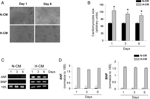
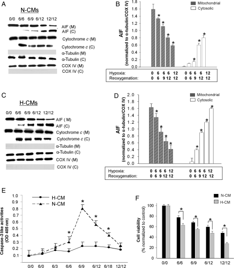
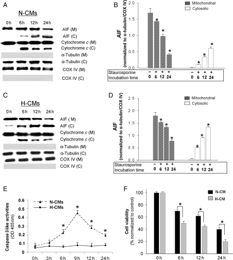
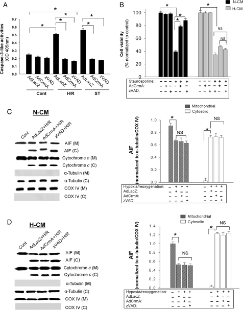
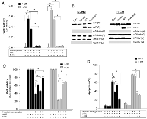
Methods and results: Hypertrophic cardiomyocyte (H-CM) cultures were prepared from the hearts of Dahl salt-sensitive rats fed a high salt diet. Apoptotic stimulation induced by hypoxia/reoxygenation or staurosporine (1 microM) enhanced AIF release in H-CMs compared with non-hypertrophic cardiomyocytes (N-CMs). Caspase inhibition using zVAD.fmk (25 microM) or overexpression of CrmA using recombinant adenovirus only partially protected N-CMs from apoptosis (63 +/- 0.93%) and provided no significant protection against apoptosis in hypertrophic cells (23 +/- 1.03%). On the other hand, poly-ADP-ribose polymerase inhibition using 4-AN (20 microM) during apoptotic stimulation blocked the release of AIF from mitochondria and significantly improved cell viability in hypertrophied cardiomyocytes (74 +/- 1.18%).

Conclusion: A caspase-dependent, apoptotic pathway is important for N-CM death, whereas a caspase-independent, AIF-mediated pathway plays a critical role in H-CMs.


Figures

Figure 1
Figure 1
Adult rat N-CM and H-CM cultures and their stability. (A) Morphology of H-CMs and N-CMs at days 1 and 6. (B) Quantitative analysis of cardiomyocyte size at days 1 and 6. n = 6. *P < 0.05. (C) PCR analysis of hypertrophic markers, ANF and BNP, at days 1, 3, and 6 in H-CMs and N-CMs. 18S was used as an internal control. (D) Quantitative analysis of ANF (left panel) and BNP (right panel) at days 1, 3, and 6 in H-CMs and N-CMs. n = 6.
Figure 2
Figure 2
Time course of AIF and cytochrome c release from mitochondria in N-CMs and H-CMs after H/R. Western blot analysis of subcellular fractions in N-CMs (A) and H-CMs (C) after different H/R conditions. To confirm the absence of significant contamination in subcellular fractions, we probed for COX IV and α-tubulin, which are internal controls for mitochondrial and cytosolic fractions, respectively. Quantitative analysis of mitochondrial and cytosolic AIF levels after different H/R conditions in N-CMs (B) and H-CMs (D). Cytosolic and mitochondrial AIF levels were normalized to α-tubulin and COX IV levels, respectively. n = 4, *P < 0.05 compared with the control. (E) Quantitative analysis of caspase-3-like activities in N-CMs (closed triangle) and H-CMs (closed circle) after different H/R conditions. Caspase-3-like activities were measured using the specific caspase substrate DEVD-pNa. OD, optical density. n = 6. *P < 0.05. (F) Quantitative analysis of cell viability after H/R in N-CMs (dark shaded) and H-CMs (light shaded). n = 6, *P < 0.05.
Figure 3
Figure 3
Time course of AIF and cytochrome c release from mitochondria in N-CMs and H-CMs after staurosporine treatment. Western blot analysis of subcellular fractions in N-CMs (A) and H-CMs (B) after staurosporine treatment. Quantitative analysis of mitochondrial and cytosolic AIF after staurosporine treatment in N-CMs (C) and H-CMs (D). Cytosolic and mitochondrial AIF levels were normalized to α-tubulin and COX IV levels, respectively. n = 4, *P < 0.05 compared with the control. (E) Quantitative analysis of caspase-3-like activities in N-CMs (closed triangle) and H-CMs (closed circle) after staurosporine treatment. Caspase-3-like activities were measured using the specific caspase substrate DEVD-pNa. n = 6. *P < 0.05. (F) Quantitative analysis of cell viability after staurosporine treatment in N-CMs (dark shaded) and H-CMs (light shaded). n = 6, *P < 0.05.
Figure 4
Figure 4
Effect of caspase inhibition on caspase-3-like activities and cell viability in cardiomyocytes after various apoptotic stimuli. (A) Inhibition of caspase-3-like activities induced by H/R and staurosporine using AdCrmA (100 m.o.i.) and zVAD.fmk (25 µM) in N-CMs. Caspase-3-like activities were measured after 6H/9R or 9 h exposure to staurosporine. (B) Effect of caspase inhibition on cell viability in N-CMs and H-CMs. Cell viability was assessed by MTT after 24 h exposure to 1 µM staurosporine. n = 6, *P < 0.05. (C, left panel) Western blot analysis of subcellular fractions in N-CMs with various caspase inhibitions (AdCrmA, 100 m.o.i. and zVAD.fmk, 25 µM) after 12H/12R. (C, right panel) Quantitative analysis of mitochondrial and cytosolic AIF levels with various caspase inhibitions after H/R in N-CMs. Cytosolic and mitochondrial AIF levels were normalized to α-tubulin and COX IV levels, respectively. n = 4, *P < 0.05. (D, left panel) Western blot analysis of subcellular fractions in H-CMs with various caspase inhibitions after H/R. (D, right panel) Quantitative analysis of mitochondrial and cytosolic AIF levels with various caspase inhibitions after H/R in H-CMs. Cytosolic and mitochondrial AIF levels were normalized to α-tubulin and COX IV levels, respectively. n = 4, *P < 0.05.
Figure 5
Figure 5
Effect of PARP inhibition in cardiomyocytes after various apoptotic stimuli. (A) PARP activity in N-CMs (dark shaded) and H-CMs (light shaded) after different apoptotic stimuli. n = 6, *P < 0.05. (B) Western blot analysis of subcellular fractions in N-CMs (left panel) and H-CMs (right panel) with PARP inhibition using 4-AN (20 µM) after H/R. (C and D) Quantitative analysis of cell viability (C) and apoptosis (D) after caspase inhibition (zVAD.fmk, 25 µM), PARP inhibition (4-AN, 20 µM), or both. Cell viability was measured by MTT assay and apoptosis was measured using annexin V staining. n = 6, *P < 0.05.
Figure 6
Figure 6
Expression of factors involved in caspase-independent apoptosis. (A) Representative western blots of various endogenous factors that modulate caspase-independent apoptosis in N-CMs and H-CMs. GAPDH is used as a loading control. (B) Quantitative analysis of these factors in N-CMs and H-CMs. Protein expressions were normalized to GAPDH expression. n = 4, *P < 0.05.

Comment in

References

    1. Gottlieb RA, Burleson KO, Kloner RA, Babior BM, Engler RL. Reperfusion injury induces apoptosis in rabbit cardiomyocytes. J Clin Invest. 1994;94:1621–1628. - PMC - PubMed
    1. Kang PM, Izumo S. Apoptosis and heart failure: a critical review of the literature. Circ Res. 2000;86:1107–1113. - PubMed
    1. Kang PM, Izumo S. Apoptosis in heart: basic mechanisms and implications in cardiovascular diseases. Trends Mol Med. 2003;9:177–182. - PubMed
    1. Abraham MC, Shaham S. Death without caspases, caspases without death. Trends Cell Biol. 2004;14:184–193. - PubMed
    1. Cande C, Cecconi F, Dessen P, Kroemer G. Apoptosis-inducing factor (AIF): key to the conserved caspase-independent pathways of cell death? J Cell Sci. 2002;115:4727–4734. - PubMed

Publication types

Substances