. 2009 Oct;297(4):L687-97.
doi: 10.1152/ajplung.90509.2008. Epub 2009 Jul 24.

A paradoxical protective role for the proinflammatory peptide substance P receptor (NK1R) in acute hyperoxic lung injury

Affiliations

A paradoxical protective role for the proinflammatory peptide substance P receptor (NK1R) in acute hyperoxic lung injury

Marwan Dib et al. Am J Physiol Lung Cell Mol Physiol. 2009 Oct.

Abstract

The neuropeptide substance P manifests its biological functions through ligation of a G protein-coupled receptor, the NK1R. Mice with targeted deletion of this receptor reveal a preponderance of proinflammatory properties resulting from ligand activation, demonstrating a neurogenic component to multiple forms of inflammation and injury. We hypothesized that NK1R deficiency would afford a similar protection from inflammation associated with hyperoxia. Counter to our expectations, however, NK1R-/- animals suffered significantly worse lung injury compared with wild-type mice following exposure to 90% oxygen. Median survival was shortened to 84 h for NK1R-/- mice from 120 h for wild-type animals. Infiltration of inflammatory cells into the lungs was significantly increased; NK1R-/- animals also exhibited increased pulmonary edema, hemorrhage, and bronchoalveolar lavage fluid protein levels. TdT-mediated dUTP nick end labeling (TUNEL) staining was significantly elevated in NK1R-/- animals following hyperoxia. Furthermore, induction of metallothionein and Na(+)-K(+)-ATPase was accelerated in NK1R-/- compared with wild-type mice, consistent with increased oxidative injury and edema. In cultured mouse lung epithelial cells in 95% O(2), however, addition of substance P promoted cell death, suggesting the neurogenic component of hyperoxic lung injury is mediated by additional mechanisms in vivo. Release of bioactive constituents including substance P from sensory neurons results from activation of the vanilloid receptor, TRPV1. In mice with targeted deletion of the TRPV1 gene, acute hyperoxic injury is attenuated relative to NK1R-/- animals. Our findings thus reveal a major neurogenic mechanism in acute hyperoxic lung injury and demonstrate concerted actions of sensory neurotransmitters revealing significant protection for NK1R-mediated functions.


Figures

Fig. 1.
Fig. 1.
Mortality of neurokinin-1 (NK1) receptor (NK1R) −/− mice is significantly greater than wild-type (WT) animals following exposure to hyperoxia. A: NK1R−/− mice on the C57BL/6 background and wild-type C57BL/6 animals of 8–12 wk were exposed to 90% O2-10% N2, and monitored for mortality. NK1R−/− animals began to die after 72 h, and 100% were dead by 96 h. Initial mortality of wild-type mice was observed at 96 h, with 100% dead by 144 h (different at P < 0.0001; n = 10–16 mice per group). B: posthyperoxia mortality was determined by exposing NK1R−/− and wild-type mice to 90% O2 for 72 h followed by return to room air. NK1R−/− mice began to die within 24 h following 72-h hyperoxia, and mortality reached 70% by 96 h compared with none for the wild-type animals (different at P < 0.005; n = 10 mice per group).
Fig. 2.
Fig. 2.
Microscopic evaluation reveals increased tissue damage in NK1R−/− mouse lungs following hyperoxia relative to wild-type animals. Photomicrographs of NK1R−/− and wild-type mouse lungs exposed to 90% O2 for 72 h are shown. The epithelial injury (*), inflammatory cell infiltrate (▴), increased alveolar thickness (↑), and entrapped red blood cells (▵) characteristic of hyperoxic injury are all significantly more pronounced in NK1R−/− mice compared with wild-type animals. Representative of 6–8 mice per group. Hematoxylin and eosin (H&E) stain, magnification ×400.
Fig. 3.
Fig. 3.
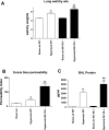
NK1R−/− mice develop more airway edema and bronchoalveolar lavage (BAL) fluid protein than wild-type animals following hyperoxia. A: lung wet/dry weights (*P < 0.0001 for hyperoxia vs. room air; **P < 0.001 for NK1R−/− vs. wild-type mice exposed to hyperoxia; n = 10 mice per group). B: increased microvascular permeability assessed by extravasation of Evans blue dye (*P < 0.05 for hyperoxia vs. room air, wild-type mice; **P < 0.0003 for NK1R−/− vs. wild-type animals after hyperoxia; n = 5–7 mice per group). C: BAL fluid protein concentration (*P < 0.0001 for hyperoxia vs. room air; **P < 0.001 for hyperoxia-exposed NK1R−/− mice vs. wild-type; n = 6–12 mice per group).
Fig. 4.
Fig. 4.
Inflammatory cell influx and hemoglobin content of NK1R−/− and wild-type mouse lungs following hyperoxia. A: the content of neutrophils in BAL fluid is elevated for wild-type but not NK1R−/− mice after exposure to hyperoxia [*P < 0.002 for wild-type mice, hyperoxia vs. room air; **no significant difference (NS) for NK1R−/− mice, hyperoxia vs. room air; n = 11–12 mice per group]. B: lung content of neutrophils determined from the tissue content of MPO (*P < 0.01 for wild-type mice, hyperoxia vs. room air; n = 6–12 mice per group; **P < 0.0001 for hyperoxia-exposed NK1R−/− vs. wild-type mice; n = 11–12 mice per group). C: lung content of hemoglobin (*P < 0.0001 for hyperoxia vs. room air; n = 6–8 mice per group; **P < 0.0005 for NK1R−/− vs. wild-type mice exposed to hyperoxia; n = 6–12 mice per group).
Fig. 5.
Fig. 5.
Hyperoxia-mediated induction of metallothionein and Na+-K+-ATPase is accelerated in NK1R−/− mice compared with wild-type. A: densitometric quantitation of Western blots for metallothionein in the lungs of wild-type and NK1R−/− mice as a function of exposure to 90% O2. Enzyme levels were increased relative to room air-exposed mice as early as 24 h after initiation of hyperoxic conditions for NK1R−/− animals (*P < 0.0001 for NK1R−/− vs. wild-type mice at 24 h; n = 6–8 mice per group). After 48- and 72-h hyperoxia exposure, both NK1R−/− and wild-type mice exhibited elevated metallothionein levels relative to room air, but the levels for NK1R−/− and wild-type animals was not distinguishable (**P < 0.0005 at 24 h, ***P < 0.001 at 72 h, hyperoxia vs. room air, NS for NK1R−/− vs. wild-type mice in hyperoxia; n = 6–8 mice per group). Data are expressed as the percent ± SE of room air-exposed animals. B: representative Western blot of metallothionein extracted from mouse lungs as a function of exposure to hyperoxia. Lung homogenate protein, 25 μg per lane, was separated by SDS-PAGE and Western blotted as described in experimental procedures. The enzyme appears as the monomer and β-ME-resistant oligomers. C: Na+-K+-ATPase levels in lung homogenates were determined by Western blotting and quantitated by densitometry. Values represent the mean percent ± SE relative to room air-exposed mice (*P < 0.0001 for NK1R−/− vs. wild-type mice at 48-h hyperoxia; **P < 0.05 for wild-type mice at 72-h vs. 48-h hyperoxia; n = 5–8 mice per group). D: representative Western blot of Na+-K+-ATPase extracted from mouse lungs as a function of exposure to hyperoxia. Lung homogenate protein, 25 μg per lane, was separated by SDS-PAGE and Western blotted as described in experimental procedures.
Fig. 6.
Fig. 6.
Cytokine levels are decreased for NK1R−/− mice compared with wild-type animals following 72-h hyperoxia. ELISA assays of TNFα, IFN-γ, and IL-1β in homogenates of NK1R−/− and wild-type mouse lungs housed in room air or 90% O2 for 72 h (TNFα, *P < 0.05 for NK1R−/− vs. wild-type mice exposed to hyperoxia; IFN-γ, *P < 0.0001 for both strains, hyperoxia vs. room air, **P < 0.005 for NK1R−/− vs. wild-type mice exposed to hyperoxia; IL-1β, *P < 0.0001 for both strains, room air vs. hyperoxia, **P < 0.0001 for NK1R−/− vs. wild-type mice exposed to hyperoxia; n = 8–15 mice per group).
Fig. 7.
Fig. 7.
NK1R−/− mice exhibit greater evidence of apoptosis compared with wild-type animals after exposure to hyperoxia. A: representative TdT-mediated dUTP nick end labeling (TUNEL) staining of sections of lung tissues from wild-type and NK1R−/− mice housed in room air or 90% O2 for 72 h. Cobalt-enhanced peroxidase staining (black) indicates apoptotic cells (representative of 3 animals per group). Sections were counterstained with nuclear fast red, magnification ×400. B: TUNEL-positive nuclei were enumerated from 20 randomly chosen fields, and 2,000 nuclei per lung were counted. Data are presented as the percent positive cells (*P < 0.05 for wild-type mice, hyperoxia vs. room air; **P < 0.0001 for NK1R−/− vs. wild-type following 72-h hyperoxia; n = 3 mice per group). C: enzymatic activity of caspase-3 in lung tissues from mice housed in room air or 90% O2 for 72 h. Equal amounts of protein were assessed colorimetrically for caspase-3 activity by reacting with N-acetyl-Asp-Glu-Val-Asp-p-nitroanilide (Ac-DEVD-pNA) as described in experimental procedures. Data are expressed as the means ± SE picomoles per minute per milligram protein (*P < 0.0001 for both strains, hyperoxia vs. room air; **NS between hyperoxia-exposed NK1R−/− and wild-type mice; n = 4–14 mice per group).
Fig. 8.
Fig. 8.
Cytotoxicity of substance P in NK1R-transfected mouse lung epithelial cells cultured in 95% O2. Murine lung epithelial MLE-12 cells stably transfected with the human NK1R were grown in room air with 5% CO2 or 95% O2-5% CO2 for 24 h in the presence or absence of 1 μM substance P (SP). Cells were harvested, stained with Alexa Fluor 488 annexin V and propidium iodide, and analyzed by flow cytometry. The percent of gated cells in each quadrant is indicated. Representative of 3 independent experiments.
Fig. 9.
Fig. 9.
Survival of transient receptor potential vanilloid type 1 (TRPV1) −/− mice in hyperoxia is increased relative to NK1R−/− animals and indistinguishable from wild-type mice. Mice of the genotypes indicated were placed in 90% O2-10% N2 and monitored for mortality. Survival of TRPV1 mice was significantly increased compared with NK1R−/− animals (P < 0.0001; n = 12 mice per group) but not different from wild-type mice (P = NS; n = 12 mice per group).

Similar articles

Cited by

References

    1. Asehnoune K, Strassheim D, Mitra S, Kim JY, Abraham E. Involvement of reactive oxygen species in Toll-like receptor 4-dependent activation of NF-kappa B. J Immunol 172: 2522–2529, 2004 - PubMed
    1. Barazzone C, Horowitz S, Donati YR, Rodriguez I, Piguet PF. Oxygen toxicity in mouse lung: pathways to cell death. Am J Respir Cell Mol Biol 19: 573–581, 1998 - PubMed
    1. Barbato JC, Catanescu O, Murray K, DiBello PM, Jacobsen DW. Targeting of metallothionein by l-homocysteine: a novel mechanism for disruption of zinc and redox homeostasis. Arterioscler Thromb 27: 49–54, 2007 - PMC - PubMed
    1. Barker GF, Manzo ND, Cotich KL, Shone RK, Waxman AB. DNA damage induced by hyperoxia: quantitation and correlation with lung injury. Am J Respir Cell Mol Biol 35: 277–288, 2006 - PMC - PubMed
    1. Bill A, Stjernschantz J, Mandahl A, Brodin E, Nilsson G. Substance P: release on trigeminal nerve stimulation, effects in the eye. Acta Physiol Scand 106: 371–373, 1979 - PubMed

Publication types

MeSH terms

LinkOut - more resources