. 2009 Oct;191(19):5953-63.
doi: 10.1128/JB.00647-09. Epub 2009 Jul 24.

The pgaABCD locus of Acinetobacter baumannii encodes the production of poly-beta-1-6-N-acetylglucosamine, which is critical for biofilm formation

Affiliations

The pgaABCD locus of Acinetobacter baumannii encodes the production of poly-beta-1-6-N-acetylglucosamine, which is critical for biofilm formation

Alexis H K Choi et al. J Bacteriol. 2009 Oct.

Abstract

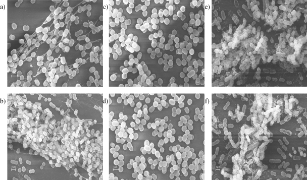
We found that Acinetobacter baumannii contains a pgaABCD locus that encodes proteins that synthesize cell-associated poly-beta-(1-6)-N-acetylglucosamine (PNAG). Both a mutant with an in-frame deletion of the pga locus (S1Deltapga) and a transcomplemented strain (S1Deltapga-c) of A. baumannii were constructed, and the PNAG production by these strains was compared using an immunoblot assay. Deleting the pga locus resulted in an A. baumannii strain without PNAG, and transcomplementation of the S1Deltapga strain with the pgaABCD genes fully restored the wild-type PNAG phenotype. Heterologous expression of the A. baumannii pga locus in Escherichia coli led to synthesis of significant amounts of PNAG, while no polysaccharide was detected in E. coli cells harboring an empty vector. Nuclear magnetic resonance analysis of the extracellular polysaccharide material isolated from A. baumannii confirmed that it was PNAG, but notably only 60% of the glucosamine amino groups were acetylated. PCR analysis indicated that all 30 clinical A. baumannii isolates examined had the pga genes, and immunoblot assays indicated that 14 of the 30 strains strongly produced PNAG, 14 of the strains moderately to weakly produced PNAG, and 2 strains appeared to not produce PNAG. Deletion of the pga locus led to loss of the strong biofilm phenotype, which was restored by complementation. Confocal laser scanning microscopy studies combined with COMSTAT analysis demonstrated that the biovolume, mean thickness, and maximum thickness of 16-h and 48-h-old biofilms formed by wild-type and pga-complemented A. baumannii strains were significantly greater than the biovolume, mean thickness, and maximum thickness of 16-h and 48-h-old biofilms formed by the S1Deltapga mutant strain. Biofilm-dependent production of PNAG could be an important virulence factor for this emerging pathogen that has few known virulence factors.


Figures

FIG. 1.
FIG. 1.
(A) Immunoblot analysis of PNAG production by A. baumannii wild-type strain S1, mutant S1Δpga, and complemented mutant S1Δpga-c using goat antibodies to dPNAG. Wild-type strain S1 and complemented mutant S1Δpga-c extracts were used at dilutions of 1:2,000, 1:4,000, and 1:8,000 (first and fourth lanes), whereas the extract from the S1Δpga sample was diluted 1:5, 1:50, and 1:500 (third lane). PNAG extracts from wild-type strain S1 and complemented mutant S1Δpga-c were digested with 50 μg/ml dispersin B (DspB) for 1 h and spotted onto the membrane (second and fifth lanes, respectively). (B) A. baumannii S1, S1Δpga, and S1Δpga-c grown on Congo red agar plates, showing differences in the colony color (red versus white) associated with production of PNAG. (C) Immunoblot analysis of PNAG production by E. coli Top10 cells harboring control plasmid pCR-XL-TOPO (first lane) or pPga containing the entire pga locus from A. baumannii S1. The dilutions of the extracts from cells harboring the control plasmid used were 1:5, 1:25, 1:50, and 1:250 and the dilutions of the extracts from cells harboring plasmid pPGA used were 1:125 and 1:250 (third lane). Extracts from cells containing either the control plasmid or pPga were treated with 50 μg/ml dispersin B for 1 h (second and fourth lanes, respectively). Immunoblotting was performed using goat antibodies specific for PNAG.
FIG. 2.
FIG. 2.
SEM of A. baumannii S1 (a and b), S1Δpga (c and d), and S1Δpga-c (e and f) at magnifications of ×5,000 and ×7,500. Bacteria adhering to coverslips were fixed, treated, and observed by SEM.
FIG. 3.
FIG. 3.
Immunoelectron microscopy: images of A. baumannii probed first with rabbit antiserum raised against dPNAG conjugated to diphtheria toxoid and then with gold-labeled donkey anti-rabbit IgG. A. baumannii S1 (a and b), S1Δpga (c and d), and S1Δpga-c (e and f) were examined at a magnification of ×13,000.
FIG. 4.
FIG. 4.
Biofilm formation by A. baumannii S1, S1Δpga, and S1Δpga-c. (A) Quantitative biofilm formation on glass surfaces by strains incubated in LB containing 1% glucose with vigorous shaking at 37°C. The bars indicate the means for 16 tubes from four independent experiments. The error bars indicate the standard errors of the means. Asterisks indicate significant differences (*, P < 0.05 [t test; n = 4]; **, P < 0.01 [t test; n = 4]). (B) Biofilms formed by A. baumannii S1 (left tube), S1Δpga (middle tube), and S1Δpga-c (right tube).
FIG. 5.
FIG. 5.
CLSM images of A. baumannii S1, S1Δpga, and S1Δpga-c biofilms after 16 h (top panels) and 48 h (lower panels) of growth. The images are maximum projections or reconstructed confocal stacks consisting of a series of xy (center), yz (left), and xz (bottom) sections. A representative CSLM image for each sample is shown.
FIG. 6.
FIG. 6.
Immunoblot analysis of PNAG production by 30 clinical isolates of A. baumannii. Extracts from strains S1 and S1Δpga were used as positive (+) and negative (−) controls, respectively. PNAG extracts were probed with goat sera raised to dPNAG.
FIG. 7.
FIG. 7.
One-dimensional (top panel) and two-dimensional (bottom panel) NMR spectra of extracts from A. baumannii S1Δpga-c identifying both acetylated glucosamines in PNAG (spots a to g) and deacetylated glucosamines in PNAG (spots h and i). The COSY spectrum cross-peaks are labeled as follows: spot a, β-GlcNAc H-1/H-2 (4.60/3.78 ppm); spot b, β-GlcNAc H-2/H-3 (3.78/3.61 ppm); spot c, β-GlcNAc H-3/H-4 (3.61/3.41 ppm); spot d, β-GlcNAc H-4/H-5 (3.41/3.63 ppm); spot e, β-GlcNAc H-5/H-6 (3.63/3.81 ppm); spot f, β-GlcNAc H-5/H-6′ (3.63/4.24 ppm); spot g, β-GlcNAc H-6/H-6′ (3.81/4.24 ppm); spot h, β-GlcNH2 H-1/H-2 (4.49/2.72 ppm); and spot i, β-GlcNH2 H-2/H-3 (2.72/3.43 ppm).

Similar articles

Cited by

References

    1. Ausubel, M., R. Brent, R. E. Kingston, D. D. Moore, J. G. Seidman, J. A. Smith, and K. Struhl (ed.). 1989. Current protocols in molecular biology. Greene Publishing Associates and Wiley-Interscience, New York, NY.
    1. Barbe, V., D. Vallenet, N. Fonknechten, A. Kreimeyer, S. Oztas, L. Labarre, S. Cruveiller, C. Robert, S. Duprat, P. Wincker, L. N. Ornston, J. Weissenbach, P. Marliere, G. N. Cohen, and C. Medigue. 2004. Unique features revealed by the genome sequence of Acinetobacter sp. ADP1, a versatile and naturally transformation competent bacterium. Nucleic Acids Res. 32:5766-5779. - PMC - PubMed
    1. Cerca, N., T. Maira-Litran, K. K. Jefferson, M. Grout, D. A. Goldmann, and G. B. Pier. 2007. Protection against Escherichia coli infection by antibody to the Staphylococcus aureus poly-N-acetylglucosamine surface polysaccharide. Proc. Natl. Acad. Sci. USA 104:7528-7533. - PMC - PubMed
    1. Choi, C. H., E. Y. Lee, Y. C. Lee, T. I. Park, H. J. Kim, S. H. Hyun, S. A. Kim, S. K. Lee, and J. C. Lee. 2005. Outer membrane protein 38 of Acinetobacter baumannii localizes to the mitochondria and induces apoptosis of epithelial cells. Cell. Microbiol. 7:1127-1138. - PubMed
    1. Cramton, S. E., C. Gerke, N. F. Schnell, W. W. Nichols, and F. Gotz. 1999. The intercellular adhesion (ica) locus is present in Staphylococcus aureus and is required for biofilm formation. Infect. Immun. 67:5427-5433. - PMC - PubMed

Publication types

MeSH terms

Associated data

LinkOut - more resources