. 2009 Sep;191(18):5706-16.
doi: 10.1128/JB.00525-09. Epub 2009 Jul 24.

Feedback control of DnaA-mediated replication initiation by replisome-associated HdaA protein in Caulobacter

Affiliations

Feedback control of DnaA-mediated replication initiation by replisome-associated HdaA protein in Caulobacter

Justine Collier et al. J Bacteriol. 2009 Sep.

Abstract

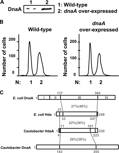
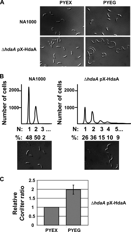
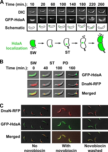
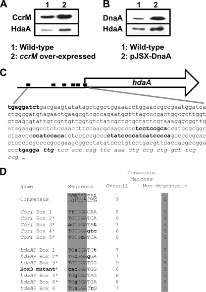
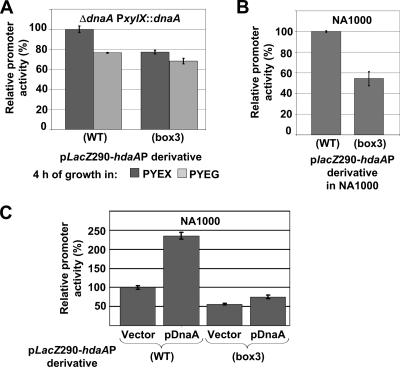
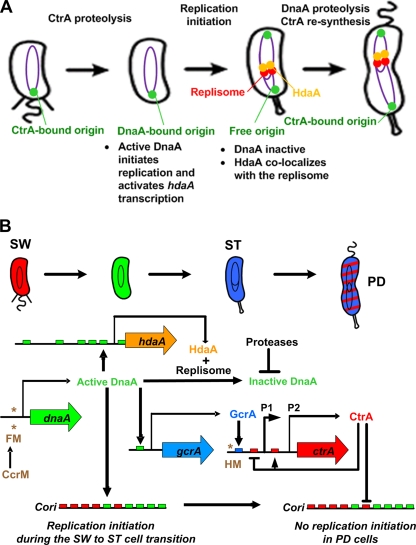
Chromosome replication in Caulobacter crescentus is tightly regulated to ensure that initiation occurs at the right time and only once during the cell cycle. The timing of replication initiation is controlled by both CtrA and DnaA. CtrA binds to and silences the origin. Upon the clearance of CtrA from the cell, the DnaA protein accumulates and allows loading of the replisome at the origin. Here, we identify an additional layer of replication initiation control that is mediated by the HdaA protein. In Escherichia coli, the Hda protein inactivates DnaA after replication initiation. We show that the Caulobacter HdaA homologue is necessary to restrict the initiation of DNA replication to only once per cell cycle and that it dynamically colocalizes with the replisome throughout the cell cycle. Moreover, the transcription of hdaA is directly activated by DnaA, providing a robust feedback regulatory mechanism that adjusts the levels of HdaA to inactivate DnaA.


Figures

FIG. 1.
FIG. 1.
The temporal control of dnaA transcription is not necessary for the correct timing of the initiation of DNA replication. (A) DnaA accumulates to high levels in cells bearing a chromosomal copy of dnaA that is transcribed from an inducible xylX promoter (dnaA overexpressed, strain JC249). Cells were grown for 4 h in PYE medium plus 0.2% glucose and 0.3% xylose (PYEGX medium), and then immunoblotting of the cell extracts was performed using antibodies to DnaA. (B) Cells from strain JC249 do not overinitiate DNA replication. Cells were grown for 4 h in PYEGX medium and then treated with rifampin for 3 h. Cells were fixed and stained with Vybrant DyeCycle orange, before analyzing their DNA content by flow cytometry. The horizontal axis indicates the number of complete chromosomes. (C) Regions of homology between the E. coli DnaA and Hda proteins and the Caulobacter DnaA and HdaA proteins are shown. The amino acid positions are shown in gray. Identities and similarities (in parentheses) between protein regions are indicated by percent values. DnaA domains I, II, III, and IV are indicated (44). DnaA domain III includes the AAA+ motifs for the ATPase activity of DnaA.
FIG. 2.
FIG. 2.
The Caulobacter hdaA gene is required for cell division and for the temporal control of the initiation of DNA replication. (A) Nomarski DIC microscopy images of NA1000 (wild-type) and JC353 (ΔhdaA pX-HdaA) cells grown in PYE medium plus xylose (PYEX; hdaA expressed) or PYE medium plus glucose (PYEG; hdaA not expressed) at all times. (B) Flow cytometry analyses of the NA1000 and JC353 strains grown for four generations in PYEG medium. Cells were treated with rifampin for 3 h prior to fixing and staining with Vybrant DyeCycle orange. The horizontal axis indicates the number of complete chromosomes. The percentages correspond to the proportion of cells containing the indicated number of chromosomes per cell. Nomarski DIC microscopy images of NA1000 and JC353 cells at the time of rifampin addition are shown in the bottom panels. (C) The JC353 strain was grown in PYEX or PYEG medium at all times, and samples were harvested to quantify the copy numbers of the chromosomal Cori and ter sites in a Southern hybridization experiment using specific probes. The ratio of the Cori sites to ter sites in cells grown in PYEX medium is defined as 1.0, and the relative value for cells grown in PYEG medium is shown. Results are the averages of data from independently duplicated experiments. The error bar indicates the standard deviation.
FIG. 3.
FIG. 3.
HdaA dynamically colocalizes with the replisome. (A) Time-lapse fluorescence microscopy analysis of GFP-HdaA-expressing cells. Strain JC208 was cultivated in M2G medium plus 0.3% xylose (M2GX) for 3 h prior to synchronization of the culture. Swarmer cells were isolated and placed on a thin layer of agarose containing nutrients, and images of the same cells were acquired at the indicated time points as the cells progressed through their cell cycle. Cell division occurs at ∼280 min under these growth conditions. DIC images, fluorescence images, and schematics of the same cells are shown. White arrows indicate cells with two closely spaced GFP-HdaA foci. The green color in the schematics indicates the intracellular position of GFP-HdaA. SW, swarmer cell; ST, stalked cell. (B) Time-lapse fluorescence microscopy analysis of GFP-HdaA and DnaN-RFP colocalization. Strain JC388 was cultivated in M2GX medium for 3 h prior to synchronization of the culture. Swarmer cells were isolated and placed on a thin layer of agarose containing nutrients, and images of the same cells were acquired at the indicated time points. Top panels, GFP-HdaA (green) over DIC image; middle panels, DnaN-RFP (red) over DIC image; bottom panels, GFP-HdaA and DnaN-RFP over DIC image to show colocalization of the fluorescence markers (yellow). SW, swarmer cell; ST, stalked cell; PD, predivisional cell. (C) Strain JC388 was synchronized, and novobiocin was added at the late stalked-cell stage (65 min into the cell cycle). Some cells were washed and resuspended in fresh M2GX medium 42 min later. Top row, DnaN-RFP (red) over DIC; middle row, GFP-HdaA (green) over DIC; bottom row, DnaN-RFP and GFP-HdaA (colocalization in yellow) over DIC; left column, untreated stalked cells 65 min into the cell cycle; middle column, cells 12 min after the addition of novobiocin; right column, cells treated with novobiocin for 42 min, washed, and incubated in fresh medium for 10 min.
FIG. 4.
FIG. 4.
DnaA promotes HdaA accumulation and the hdaA promoter region contains multiple putative DnaA boxes. (A) Strain JC362, with an extra chromosomal copy of ccrM under the control of the constitutive lacZ promoter (ccrM overexpressed) and the wild-type strain, with a single cell cycle-regulated copy of ccrM, were grown in PYE medium and used to perform immunoblotting using antibodies raised against CcrM and HdaA. (B) A wild-type strain, with or without pJSX-DnaA, was grown in PYE medium plus 0.2% glucose and 0.3% xylose (PYEGX) for 4 h and used to perform immunoblotting using antibodies raised against DnaA and HdaA. (C) The nucleotide sequence of the hdaA promoter region (345 nucleotides) is shown. The nucleotides corresponding to the six putative DnaA boxes are in bold. Italics indicate the beginning of the hdaA coding sequence. The black boxes in the schematic above indicate the DnaA boxes. (D) Consensus sequence for DnaA boxes in Caulobacter (17) and the putative DnaA boxes within the Cori (30) and the hdaA promoter. Reverse-complement sequences are presented for boxes marked with asterisks. Nucleotide positions differing from the consensus are denoted in lowercase bold lettering. The six nondegenerate nucleotides of the consensus DnaA box are highlighted in gray. The number of nucleotide matches to the overall and nondegenerate consensus DnaA box are shown for each sequence. The sequence of the mutated hdaAP(box3) is displayed, with lowercase bold letters indicating nucleotide changes compared to the wild type.
FIG. 5.
FIG. 5.
DnaA directly activates the transcription of hdaA. (A) The graph shows the relative β-galactosidase activities from placZ290-hdaAP(WT) and placZ290-hdaAP(box3) in a GM2471 (ΔdnaA PxylX::dnaA) strain upon the depletion of DnaA 4 h after a shift from PYE medium plus xylose (PYEX) to PYE medium plus glucose (PYEG). (B) The graph shows the relative β-galactosidase activities from placZ290-hdaAP(WT) and placZ290-hdaAP(box3) in an NA1000 (WT) strain grown in PYE medium. Activities in Miller units were normalized so that the activity of hdaAP(WT) equals 100% in PYE (NA1000) or PYEX (GM2471) medium to facilitate comparison. (C) The graph shows the relative β-galactosidase activities from placZ290-hdaAP(WT) and placZ290-hdaAP(box3) in an NA1000 strain containing the DnaA-overexpressing plasmid pJSX-DnaA or the empty control vector pJS14 grown in PYEX medium for 4 h. Activities in Miller units were normalized so that the activity of hdaAP(WT) equals 100% in NA1000 containing pJS14 to facilitate comparison. Error bars indicate the standard deviations.
FIG. 6.
FIG. 6.
Model for the temporal control of DNA replication initiation in Caulobacter. (A) A schematic of the beginning of the Caulobacter cell cycle is shown. Purple theta structures inside the cells indicate replicating DNA. The single origin of replication (green focus) in swarmer cells is bound to CtrA, which represses the initiation of DNA replication. During the swarmer-to-stalked cell transition, CtrA is rapidly degraded by the ClpXP protease, and active DnaA binds to the origin to initiate DNA replication. The replisome (red foci), associated with HdaA (orange foci), replicates the chromosome and inactivates DnaA once DNA replication is ongoing. CtrA reaccumulates in predivisional cells and binds to the origin to prevent more replication initiation events. (B) A schematic of the beginning of the Caulobacter cell cycle is shown. Red indicates CtrA accumulation, green indicates DnaA accumulation, and blue indicates GcrA accumulation. SW, swarmer cell; ST, stalked cell; PD, predivisional cell. DnaA is synthesized in swarmer cells, when the dnaA promoter is in the fully methylated state (FM, two asterisks). New molecules of DnaA initiate DNA replication and activate the transcription of gcrA and hdaA by directly binding to DnaA boxes (green boxes). Once the replisome is assembled, the replisome-HdaA complex inhibits the initiation of DNA replication, probably by a mechanism similar to the RIDA mechanism in E. coli, and the DnaA protein is degraded by a protease (15) to prevent more initiation events in stalked cells. Soon after the initiation of DNA replication, the dnaA and the ctrA genes are duplicated by the passage of the replication fork and therefore hemimethylated (HM, asterisk). Transcription from the hemimethylated dnaA gene is shut down, while transcription from the hemimethylated ctrA gene is turned on by the binding of GcrA to the ctrA P1 promoter (blue box). Accumulation of CtrA in early predivisional cells then contributes to the inactivation of replication initiation by directly binding to the CtrA sites in the Cori (red boxes), yielding a robust replication control system.

Similar articles

Cited by

References

    1. Banack, T., N. Clauson, N. Ogbaa, J. Villar, D. Oliver, and W. Firshein. 2005. Overexpression of the Hda DnaA-related protein in Escherichia coli inhibits multiplication, affects membrane permeability, and induces the SOS response. J. Bacteriol. 1878507-8510. - PMC - PubMed
    1. Bastedo, D. P., and G. T. Marczynski. 2009. CtrA response regulator binding to the Caulobacter chromosome replication origin is required during nutrient and antibiotic stress as well as during cell cycle progression. Mol. Microbiol. 72139-154. - PubMed
    1. Brendler, T., A. Abeles, and S. Austin. 1995. A protein that binds to the P1 origin core and the oriC 13mer region in a methylation-specific fashion is the product of the host seqA gene. EMBO J. 144083-4089. - PMC - PubMed
    1. Collier, J., H. H. McAdams, and L. Shapiro. 2007. A DNA methylation ratchet governs progression through a bacterial cell cycle. Proc. Natl. Acad. Sci. USA 10417111-17116. - PMC - PubMed
    1. Collier, J., S. R. Murray, and L. Shapiro. 2006. DnaA couples DNA replication and the expression of two cell cycle master regulators. EMBO J. 25346-356. - PMC - PubMed

Publication types

MeSH terms

LinkOut - more resources