. 2009 Aug;136(16):2767-77.
doi: 10.1242/dev.034454.

The apicobasal polarity kinase aPKC functions as a nuclear determinant and regulates cell proliferation and fate during Xenopus primary neurogenesis

Affiliations

The apicobasal polarity kinase aPKC functions as a nuclear determinant and regulates cell proliferation and fate during Xenopus primary neurogenesis

Nitin Sabherwal et al. Development. 2009 Aug.

Abstract

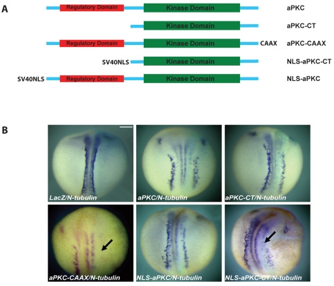
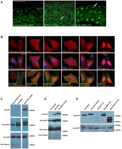
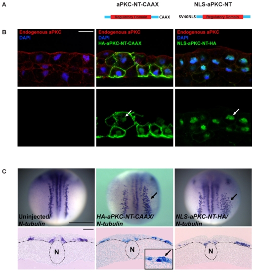
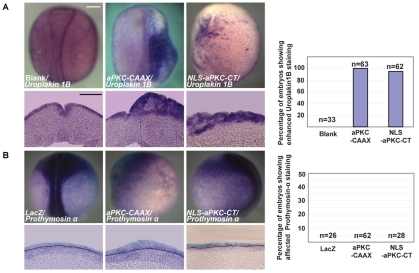
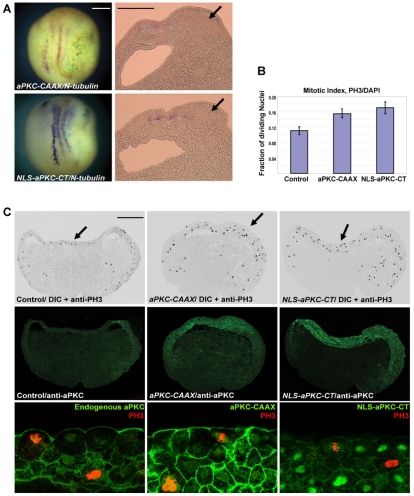
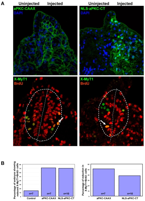
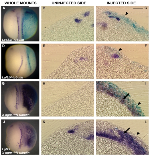
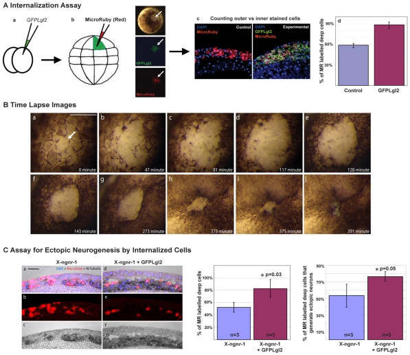
During neurogenesis in Xenopus, apicobasally polarised superficial and non-polar deep cells take up different fates: deep cells become primary neurons while superficial cells stay as progenitors. It is not known whether the proteins that affect cell polarity also affect cell fate and how membrane polarity information may be transmitted to the nucleus. Here, we examine the role of the polarity components, apically enriched aPKC and basolateral Lgl2, in primary neurogenesis. We report that a membrane-tethered form of aPKC (aPKC-CAAX) suppresses primary neurogenesis and promotes cell proliferation. Unexpectedly, both endogenous aPKC and aPKC-CAAX show some nuclear localisation. A constitutively active aPKC fused to a nuclear localisation signal has the same phenotypic effect as aPKC-CAAX in that it suppresses neurogenesis and enhances proliferation. Conversely, inhibiting endogenous aPKC with a dominant-negative form that is restricted to the nucleus enhances primary neurogenesis. These observations suggest that aPKC has a function in the nucleus that is important for cell fate specification during primary neurogenesis. In a complementary experiment, overexpressing basolateral Lgl2 causes depolarisation and internalisation of superficial cells, which form ectopic neurons when supplemented with a proneural factor. These findings suggest that both aPKC and Lgl2 affect cell fate, but that aPKC is a nuclear determinant itself that might shuttle from the membrane to the nucleus to control cell proliferation and fate; loss of epithelial cell polarity by Lgl2 overexpression changes the position of the cells and is permissive for a change in cell fate.


Figures

Fig. 1.
Fig. 1.
The effect of overexpressing aPKC on primary neurogenesis in Xenopus. (A) Schematic representation of the various aPKC constructs used in this work. SV40NLS, nuclear localisation signal from SV40; CAAX, prenylation signal. (B) mRNAs for aPKC (1 ng), aPKC-CT (0.5 ng), aPKC-CAAX (0.25-0.5 ng), NLS-aPKC (1 ng) and NLS-aPKC-CT (0.25-0.5 ng) were injected unilaterally together with lacZ mRNA (0.25 ng) and embryos were analysed by in situ hybridisation for N-tubulin expression. Only aPKC-CAAX and NLS-aPKC-CT showed significant inhibitory effects on primary neurogenesis (arrows). Scale bar: 250 μm.
Fig. 2.
Fig. 2.
Subcellular localisation and expression of aPKC constructs. (A) Immunohistochemistry using anti-aPKC antibody (green) on sections showing ectoderm of st.10.5 Xenopus embryos (apical side up) from non-injected control (left) or injection with aPKC-CAAX or NLS-aPKC-CT mRNAs. Arrows indicate nuclear staining. (B) Immunocytochemistry on HeLa cells transfected with the constructs shown and assayed with anti-aPKC (red) and anti-GFP (green) antibodies. Nuclei are blue (DAPI staining). (C) Western blot analysis with nuclear lysates from HeLa cells transfected with HA-tagged constructs as shown and assayed with anti-aPKC and anti-HA-antibodies. Endogenous aPKC shows some nuclear localisation, as shown by the anti-aPKC antibody, but HA-tagged aPKC-CAAX shows greater nuclear accumulation than HA-aPKC, as shown by the anti-HA antibody. P95 and tubulin were used as nuclear and cytoplasmic controls, respectively. (D) Western blot analysis with nuclear lysates from st.10.5 Xenopus embryos injected with HA-tagged constructs as shown and blotted with anti-HA and anti-aPKC antibodies. Both HA-tagged aPKC and aPKC-CAAX show nuclear accumulation. Lamin and tubulin were used as nuclear and cytoplasmic controls, respectively. (E) Expression analysis using whole cell lysates from HeLa cells overexpressing various aPKC constructs as shown. GFP, control for transfection and loading. Scale bars: 50 μm in A; 250 μm in B.
Fig. 3.
Fig. 3.
Effects of overexpressing dominant-negative aPKC contructs on primary neurogenesis. (A) The two dominant-negative forms of aPKC used. (B,C) Xenopus embryos unilaterally injected with either HA-aPKC-NT-CAAX or NLS-aPKC-NT-HA mRNA were processed for (B) immunohistochemistry on sectioned ectoderm using anti-HA (green) and anti-aPKC (red) antibodies, or (C) for in situ hybridisation at st.16 for N-tubulin expression. Whereas HA-aPKC-NT-CAAX showed predominantly cortical staining with weak nuclear localisation, NLS-aPKC-NT-HA showed exclusively nuclear localisation (B). Overexpression of both dominant-negatives enhanced neurogenesis (C). Arrows point to nuclear staining (B), ectopic neurons (C, upper row) and ectopic neurons in superficial neuroectoderm (C, lower row, middle panel inset). Dotted lines (C, lower row) indicate the boundary between ectoderm and mesoderm. N, notochord. Scale bars: 50 μm in B; 1 mm in C, upper row; 500 μm in C, lower row.
Fig. 4.
Fig. 4.
Effects of overexpressing aPKC constructs on expression of superficial and deep layer markers. Whole-mounts and sections of st.16 Xenopus embryos showing in situ hybridisation for (A) the superficial ectodermal marker Uroplakin 1B and (B) the deep ectodermal marker Prothymosin alpha. The dotted line marks the boundary between ectoderm (above) and mesendoderm (below). Both ectodermal layers are thickened following injection of either aPKC-CAAX or NLS-aPKC-CT mRNA. n, total number for embryos analysed. Scale bars: 250 μm.
Fig. 5.
Fig. 5.
Effect of overexpressing aPKC constructs on cell proliferation. (A) Neurula stage Xenopus embryos overexpressing aPKC-CAAX and NLS-aPKC-CT showed suppression of N-tubulin expression and a thickened ectoderm (arrows). (B) Quantitation of phospho-histone H3 (PH3)-positive nuclei relative to the total number of nuclei as obtained from at least three embryos in each set and a minimum of five sections per embryo. (C) Sections of st.10.5 embryos injected with aPKC-CAAX and NLS-aPKC-CT and co-immunostained with anti-PH3 (black in DIC image, top row, and red in bottom row) and anti-aPKC (green, in middle and bottom rows) antibodies. Sections show multilayered, thickened ectoderm and increased PH3 staining in both layers of the ectoderm. The bottom row shows the middle row at higher magnification, with anti-aPKC and anti-PH3. Scale bars: 250 μm in A; 50 μm in C.
Fig. 6.
Fig. 6.
Effect of overexpressing aPKC-CAAX or NLS-aPKC-CT on the neuronal population born during gastrulation. Xenopus embryos were unilaterally injected at the 2-cell stage with either aPKC-CAAX or NLS-aPKC-CT, BrdU saturated at st.10.5 and immunostained for BrdU and for the neuronal differentiation marker X-MyT1 on sections of tailbud at st.25. (A) Top row shows anti-aPKC staining (green), with nuclei in blue (DAPI), to distinguish between injected and non-injected sides for aPKC-CAAX and NLS-aPKC-CT injections. Bottom row shows adjacent sections co-stained for BrdU (red) and X-MyT1 (green). Neural progenitors are positive for BrdU only and appear red, neurons born after BrdU labelling are co-labelled and appear orange (arrow), and neurons born before st.10.5 (BrdU saturation) appear green. The number of neurons born before st.10.5 (green) is reduced after overexpression of either aPKC construct. The dashed lines in circle delineate the neural tube and the straight dashed lines mark the midline of the neural tube. (B) Quantification of the results from A.
Fig. 7.
Fig. 7.
Effect of Lgl2 overexpression on primary neurogenesis. Overexpression of Lgl2 (1 ng) had no effect on expression of the primary neuron marker N-tubulin (n=35) (D-F) by comparison with control embryos injected with lacZ mRNA only (0.25 ng, n=32) (A-C). But when Lgl2 was co-expressed with the proneurogenic factor X-ngnr-1 (0.25 ng, n=26) (J-L), ectopic primary neurons were found in a position intermediate between the superficial and deep layers (L, arrow), whereas embryos injected with X-ngnr-1 alone (n=41) (G-I) showed ectopic neurogenesis only in the deep layer of neuroectoderm (I, arrow). Also note the absence of lacZ staining in the superficial layer cells of embryos injected with Lgl2 and X-ngnr-1 (L, arrowhead), as compared with lacZ (C) or X-ngnr-1 (I) injected embryos, indicating the possibility of internalisation of superficial cells due to Lgl2 overexpression. Scale bars: 250 μm.
Fig. 8.
Fig. 8.
Lgl2-expressing cells become internalised and contribute to primary neurogenesis. (A) The distribution of GFPLgl2-expressing cells in the inner versus outer ectodermal layer of neurula stage Xenopus embryos was assessed by an internalisation assay (see Materials and methods for details) that involved lineage-labelling individual GFPLgl2-expressing outer cells by Micro-Ruby injection at the blastula stage (a,b). The fraction of Micro-Ruby-labelled cells in the inner layer over the total number of labelled cells was increased in GFPLgl2-injected embryos as compared with control embryos (c,d). (B) Time-lapse images of an embryo overexpressing GFPLgl2, showing early loss of apicobasal cell polarity in superficial cells (apical de-pigmentation, arrow in a) and subsequent internalisation of these cells at mid-gastrula stage (g-j). (C) To determine whether internalised cells resulting from GFPLgl2 overexpression contribute to increased neurogenesis, the Micro-Ruby-based internalisation assay was combined with in situ hybridisation for N-tubulin, as analysed in sections of st.16 neurulae from embryos that had been injected at the 2-cell stage with either X-ngnr-1 alone or in combination with GFPLgl2 (a-f). Bar charts show that the percentage of Micro-Ruby-labelled deep layer cells relative to the total number of labelled cells was increased when GFPLgl2 was co-injected and that ∼76% of these cells, on average, were also N-tubulin positive. Scale bars: 500 μm in B; 50 μm in C.

Similar articles

Cited by

References

    1. Asashima, M. and Grunz, H. (1983). Effects of inducers on inner and outer gastrula ectoderm layers of Xenopus laevis. Differentiation 23, 206-212. - PubMed
    1. Atwood, S. X. and Prehoda, K. E. (2009). aPKC phosphorylates miranda to polarize fate determinants during neuroblast asymmetric cell division. Curr. Biol. 19, 723-729. - PMC - PubMed
    1. Bellefroid, E. J., Bourguignon, C., Hollemann, T., Ma, Q., Anderson, D. J., Kintner, C. and Pieler, T. (1996). X-MyT1, a Xenopus C2HC-type zinc finger protein with a regulatory function in neuronal differentiation. Cell 87, 1191-1202. - PubMed
    1. Betschinger, J. and Knoblich, J. A. (2004). Dare to be different: asymmetric cell division in Drosophila, C. elegans and vertebrates. Curr. Biol. 14, R674-R685. - PubMed
    1. Bourguignon, C., Li, J. and Papalopulu, N. (1998). XBF-1, a winged helix transcription factor with dual activity, has a role in positioning neurogenesis in Xenopus competent ectoderm. Development 125, 4889-4900. - PubMed

Publication types

MeSH terms