. 2009 Aug;136(16):2849-60.
doi: 10.1242/dev.035246.

Wash functions downstream of Rho and links linear and branched actin nucleation factors

Affiliations

Wash functions downstream of Rho and links linear and branched actin nucleation factors

Raymond Liu et al. Development. 2009 Aug.

Abstract

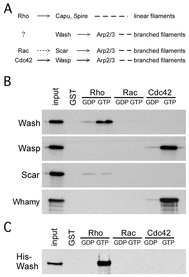
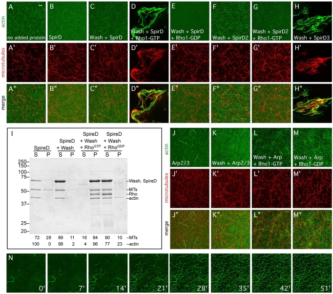
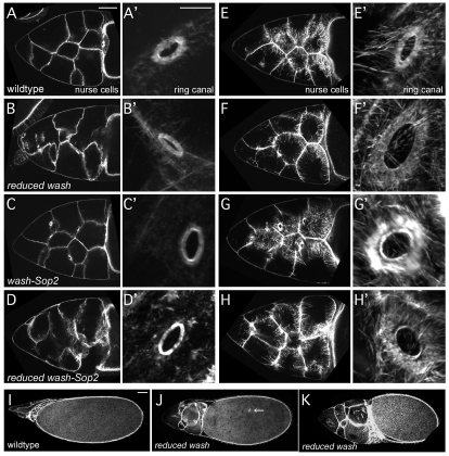
Wiskott-Aldrich Syndrome (WAS) family proteins are Arp2/3 activators that mediate the branched-actin network formation required for cytoskeletal remodeling, intracellular transport and cell locomotion. Wasp and Scar/WAVE, the two founding members of the family, are regulated by the GTPases Cdc42 and Rac, respectively. By contrast, linear actin nucleators, such as Spire and formins, are regulated by the GTPase Rho. We recently identified a third WAS family member, called Wash, with Arp2/3-mediated actin nucleation activity. We show that Drosophila Wash interacts genetically with Arp2/3, and also functions downstream of Rho1 with Spire and the formin Cappuccino to control actin and microtubule dynamics during Drosophila oogenesis. Wash bundles and crosslinks F-actin and microtubules, is regulated by Rho1, Spire and Arp2/3, and is essential for actin cytoskeleton organization in the egg chamber. Our results establish Wash and Rho as regulators of both linear- and branched-actin networks, and suggest an Arp2/3-mediated mechanism for how cells might coordinately regulate these structures.


Figures

Fig. 1.
Fig. 1.
Wash activates Arp2/3 to nucleate actin, and bundles F-actin and microtubules. (A-C) Pyrene-actin polymerization assays. Wash, Wasp and Scar VCA fragments (A) and full-length (FL) proteins (B) nucleate actin in the presence of Arp2/3. Wash nucleation activity is concentration dependent and requires Arp2/3 (C). All polymerization assays were performed a minimum of three times. (D) Coomassie-stained protein gels of full-length and VCA fragments of Wasp family proteins used in this study. (E-K″) Stabilized F-actin (1 μM; top) and MTs (1 μM; middle) were incubated with WAS family proteins and observed by confocal microscopy: (E) no protein added; (F) Wash with actin only; (G) Wash with MTs only; (H-H″) Wash; (I-I″) Wasp; (J-J″) Scar; (K-K″) Whamy. (L-T) Electron micrographs of negatively stained samples from the indicated F-actin and MT bundling/crosslinking assays above. Two examples are shown for each assay with Wash (M-T). (U,V) Quantification of F-actin and MT bundling/crosslinking efficiency of indicated reactions by low-speed pelleting assays. The crosslinking/bundling reactions described above were centrifuged to pellet actin and microtubule bundles. Supernatants (S) and pellets (P) were separated and analyzed by Coomassie staining. Percentages of protein, actin and microtubules in each fraction are given. All assays were performed a minimum of three times. Final protein concentrations: Wash, 300 nM; Wasp, 300 nM; Scar, 300 nM; Whamy, 300 nM. Scale bars: 10 μm for E-K″; 500 nm for L-T.
Fig. 2.
Fig. 2.
Wash is essential for oocyte development, functions in the Rho1/Capu/Spire pathway, and is enriched at the oocyte cortex. (A-D) Compared with wild type (A), eggs from reduced wash females have fused dorsal appendages (B,C) and a shorter egg length with no dorsal appendages (D). (E-I′) Individual frames (left) and projections (middle) from a 30-minute movie of stage 7 oocytes. Wild-type oocytes show no directed or coordinated movement of yolk granules (E), whereas oocytes from reduced wash (wash/+; wimp/+ females; F), wash RNAi (G), wash-Rho1 transheterozygous (H) and wash-capu transheterozygous (I) females undergo premature ooplasmic streaming (see Movies 1-5 in the supplementary material). The speed of yolk granules from active, swirling regions of mutant oocytes, or a random region in non-swirling wild-type oocytes are shown in units of nm/second. (J-N) α-tubulin staining of fixed wild-type stage 7 oocytes (J) show an ordered gradient of MT organization that is lost in reduced wash (K), wash RNAi (L), wash-Rho1 transheterozygous (M) and wash-capu transheterozygous (N) mutants. Anterior is to the top. Arrows in J indicate the anteroposterior gradient of microtubule organization in wild-type oocytes; arrows in K-N indicate loss of this gradient in mutant oocytes; arrowhead in K indicates a mislocalized nucleus. (O-S′) Phalloidin staining of fixed wild-type stage 7 oocytes (O) displays a band of cortical actin (enlarged in O′-S′) that is disrupted in reduced wash (P), wash RNAi (Q), wash-Rho1 transheterozygous (R) and wash-capu transheterozygous (S) mutants. (T-V) Embryos fixed and stained for Engrailed. wash/spire transheterozygous embryos display segmentation defects. (W-X′) Wash expression in the developing stage 7 (W,W′) and stage 10 (X,X′) egg chamber. Note the accumulation of Wash protein at the oocyte cortex throughout oogenesis. Scale bars: in A, 50 μm for A-D; in E, 10 μm for E-S; in O′, 2 μm for O'-S'; in T, 50 μm for T-V; in W,X, 10 μm; in W',X', 50 μm.
Fig. 3.
Fig. 3.
Wash preferentially binds active Rho but not Rac or Cdc42. (A) Summary of the previously known associations among Rho GTPases, WASP family proteins and actin nucleators. (B) GST pull-down assays with 35S-labeled in vitro translated proteins on the left and GST fusion proteins or GST alone across the top. Wash binds to Rho1GTP, but not Rac or Cdc42. By comparison, Wasp binds Cdc42GTP, whereas Scar binds directly to none of the three effectors. A fourth WASP subfamily protein in Drosophila, Whamy (CG12946), binds Cdc42GTP. (C) GST pull-down assay with purified His-Wash and GST-Rho family proteins. Wash binds directly to Rho1GTP, but not Rac or Cdc42. All assays were performed a minimum of three times.
Fig. 4.
Fig. 4.
Wash interacts with the linear actin nucleators Capu and Spire. (A,B) Diagrams of the Drosophila Wash protein and the SpirA, SpirC and SpirD protein isoforms. Protein fragments used to map protein-protein interactions are indicated, with interacting fragments highlighted in bold. (C-F) GST pull-down assays with 35S-labeled in vitro translated proteins on the left and GST fusion proteins or GST alone across the top. (C) Full-length (FL) Wash binds all Spire isoforms and itself through fragment A of Wash. (D-F) Full-length (FL) Wash binds fragment C3a of SpirC, and fragment D2 of SpirD. (G) Western blot of immunoprecipitations (IPs) from embryo extracts performed with no antibody (no Ab), a nonspecific antibody (9e10) or Wash antibody. (H-K) Western blots of IPs from embryo extracts with Wash, Rho1, no primary antibody, or with an unrelated antibody (9e10). (H) Wash is present in both Wash and Rho immunoprecipitated complexes. (I-K) Capu, SpirD and SpirC are present in Wash immunoprecipitated complexes. All assays were performed a minimum of three times.
Fig. 5.
Fig. 5.
Wash F-actin/MT bundling and crosslinking activity is regulated by Rho1, Spire and Arp2/3. (A-H″) Stabilized F-actin (1 μM, A-H) and MTs (1 μM, A′-H′) were incubated with full-length Wash and/or Rho1, Arp2/3 and Spire, and observed by confocal microscopy: (A) no protein added; (B) SpirD only; (C) Wash and SpirD; (D) Wash, SpirD and Rho1GTP; (E) Wash, SpirD and Rho1GDP; (F) Wash and SpirD2; (G) Wash, SpirD2 and Rho1GTP; (H) Wash and SpirD3. (I) Quantification of F-actin and MT bundling/crosslinking efficiency of indicated reactions (see Fig. 3; quantification of Arp2/3 reactions were not obtained owing to co-migration of Arp2/3 with actin and MT bands). (J-M″) F-actin (J-M) and microtubule bundling (J′-M′) and crosslinking assay with: (J) Arp2/3; (K) Wash and Arp2/3; (L) Wash, Arp2/3 and Rho1GTP; (M) Wash, Arp2/3 and Rho1GDP. All assays were performed a minimum of three times. Final protein concentrations were: Wash, 300 nM; Arp2/3, 600 nM; Rho1GTP, 600 nM; SpirD, 300 nM; SpirD2, 300 nM; SpirD3, 300 nM. (N) Stills from a time-lapse confocal movie of branched-actin network formation by Wash and Arp2/3 in the presence of F-actin (see Movie 6 in the supplementary material). Scale bar: 10 μm.
Fig. 6.
Fig. 6.
Wash functions with Arp2/3 to regulate actin cyto-architecture in oogenesis. (A-H′) Nurse cells (A-H) and ring canals (A′-H′) from stage 10a (A-D′) and stage 10b-11 (E-H′) egg chambers fixed and stained with phalloidin. Reduced wash (wash/+; wimp/+ females), wash-Sop2 (Sop2 wash/+ + females) and reduced wash-Sop2 (Sop2 wash/+ +; wimp/+ females) mutant oocytes (B-D,F-H) show a loss of nurse cell integrity and incomplete stress fiber formation compared with wild type (A,E). Reduced wash, wash-Sop2 and reduced wash-Sop2 mutant oocytes do not affect ring canal formation (B′-D′), but do exhibit abnormal actin organization around ring canals by stage 10b-11 (F′-H′). (I-K) Stage 14 egg chambers fixed and stained with phalloidin. Compared with wild type (I), reduced wash mutant nurse cells (J,K) retain more of their cytoplasm (incomplete dumping), and oocytes are smaller. Consistent with defects in actin cyto-architecture, ring canals separated from the nurse cell membrane can be found `floating' within the oocyte (arrow in H). Scale bars: in A, 50 μm for A-H; in A′, 10 μm for A'-H'; in I, 50 μm for I-K.
Fig. 7.
Fig. 7.
Proposed model of Rho-regulated Wash maintenance of egg chamber cyto-architecture. (A) In nurse cells, Rho activates Wash nucleation of branched actin filaments (left) providing structural integrity for nurse cells and ring canals. In the oocyte, active Rho inhibits SpirD (right), freeing Capu, SpirC and Wash to crosslink/bundle cortical microfilaments and MTs, and preventing premature ooplasmic streaming (see illustration in B). Upon an unknown signaling event during stage 10b, SpirD is released from RhoGTP inhibition, binds to Capu, SpirC and Wash, and inhibits their crosslinking/bundling activity. This initiates the formation of subcortical MT arrays (B′), leading to ooplasmic streaming. (B,B′) Illustration of signaling events depicted in A.

Similar articles

Cited by

References

    1. Bartolini, F., Moseley, J. B., Schmoranzer, J., Cassimeris, L., Goode, B. L. and Gundersen, G. G. (2008). The formin mDia2 stabilizes microtubules independently of its actin nucleation activity. J. Cell Biol. 181, 523-536. - PMC - PubMed
    1. Bement, W. M., Miller, A. L. and von Dassow, G. (2006). Rho GTPase activity zones and transient contractile arrays. BioEssays 28, 983-993. - PMC - PubMed
    1. Ben-Yaacov, S., Le Borgne, R., Abramson, I., Schweisguth, F. and Schejter, E. D. (2001). Wasp, the Drosophila Wiskott-Aldrich syndrome gene homologue, is required for cell fate decisions mediated by Notch signaling. J. Cell Biol. 152, 1-13. - PMC - PubMed
    1. Berger, S., Schafer, G., Kesper, D. A., Holz, A., Eriksson, T., Palmer, R. H., Beck, L., Klambt, C., Renkawitz-Pohl, R. and Onel, S. F. (2008). WASP and SCAR have distinct roles in activating the Arp2/3 complex during myoblast fusion. J. Cell Sci. 121, 1303-1313. - PubMed
    1. Bione, S., Sala, C., Manzini, C., Arrigo, G., Zuffardi, O., Banfi, S., Borsani, G., Jonveaux, P., Philippe, C., Zuccotti, M. et al. (1998). A human homologue of the Drosophila melanogaster diaphanous gene is disrupted in a patient with premature ovarian failure: evidence for conserved function in oogenesis and implications for human sterility. Am. J. Hum. Genet. 62, 533-541. - PMC - PubMed

Publication types

MeSH terms