. 2010 Mar;20(3):704-19.
doi: 10.1093/cercor/bhp136. Epub 2009 Jul 24.

Forelimb muscle representations and output properties of motor areas in the mesial wall of rhesus macaques

Affiliations

Forelimb muscle representations and output properties of motor areas in the mesial wall of rhesus macaques

Marie-Hélène Boudrias et al. Cereb Cortex. 2010 Mar.

Abstract

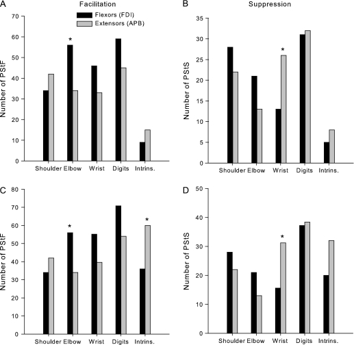
In this study, forelimb organizations and output properties of the supplementary motor area (SMA) and the dorsal cingulate motor area (CMAd) were assessed and compared with primary motor cortex (M1). Stimulus-triggered averages of electromyographic activity from 24 muscles of the forelimb were computed from layer V sites of 2 rhesus monkeys performing a reach-to-grasp task. No clear segregation of the forelimb representation of proximal and distal muscles was found in SMA. In CMAd, sites producing poststimulus effects in proximal muscles tended to be located caudal to distal muscle sites, although the number of effects was limited. For both SMA and CMAd, facilitation effects were more prevalent in distal than in proximal muscles. At an intensity of 60 microA, the mean latencies of M1 facilitation effects were 8 and 12.1 ms shorter and the magnitudes approximately 10 times greater than those from SMA and CMAd. Our results show that corticospinal neurons in SMA and CMAd provide relatively weak input to spinal motoneurons compared with the robust effects from M1. However, a small number of facilitation effects from SMA and CMAd had latencies as short as the shortest ones from M1 suggesting a minimum linkage to motoneurons as direct as that from M1.


Figures

Figure 1.
Figure 1.
Reconstruction procedures applied to the mesial wall of the cortex based on MR images and electrophysiological data. (A) Surface view of the electrode penetration maps of the left hemisphere for both monkeys. Dotted lines indicate anatomical sulci. Abbreviations: A, Anterior; ArS, arcuate sulcus; CS, central sulcus; M, medial; MID, midline; and SPS, superior precentral sulcus. Numbers 1–11 are 1 mm apart and referenced to the coronal section represented on the unfolded layer V map in (C). Tracks where ICMS produced face and mouth movements in M1 and ventral premotor area (PMv) are marked by purple–pink dots; tracks where ICMS-evoked hindlimb movements in M1 are marked by yellow dots; tracks randomly selected from the M1 forelimb area for stimulation at both 15 and 60 μA are marked by orange dotes; tracks where StTA yielded PStEs from SMA are marked by red dots; tracks more lateral along the hemisphere that intersected SMA or CMAd deep along the mesial surface and produced effects are marked by blue dots; tracks where StTA did not produce PStEs are marked by small black dots; and tracks used for surface reconstruction of the brain but where data collected were not included in this analysis are marked by small gray dots; (B) Coronal section (Monkey J, section 1) of the mesial wall and the cortical dorsal surface of SMA based on MRI image; (C) Map of unfolded layer V of the gray matter of SMA and CMAd represented in 2D coordinates.
Figure 2.
Figure 2.
Two-dimensional unfolded layer V maps of the mesial wall showing the organization of PStF and PStS at each joint (shoulder, elbow, wrist, digit, and intrinsic hand) based on StTA of EMG activity at 60 μA for each monkey. The color-coded motor representation for proximal and distal joints is shown in the figure. Map unfolding procedures are explained in Materials and Methods.
Figure 3.
Figure 3.
Maps of SMA and CMAd for 2 monkeys represented in 2D coordinates after unfolding the mesial wall of the cortex. Maps are based on PStF and PStS effects at proximal and distal joints. Effects marked with an X were not included in the SMA or CMAd data set because they were located on the border of 2 motor areas. Map unfolding procedures are explained in Materials and Methods.
Figure 4.
Figure 4.
Distribution of PStF (right) and PStS (left) obtained from 19 to 23 muscles of the forelimb for SMA (A) and CMAd (B). The dotted lines separate muscles belonging to different joints. See Materials and Methods for muscle abbreviations. The asterisk (*) on muscles FCU and ED 4,5 indicates effects from monkey J only. Because of the combination of muscles formed to produce TRI and Intrins. in monkey Y, the total number of effects in these muscles was divided by 2 and distributed equally in muscles labeled TLON and TLAT and in muscles labeled FDI and APB.
Figure 5.
Figure 5.
(A,B) Distribution of PStF and PStS from SMA in extensor and flexor muscles of the shoulder, elbow, wrist, and digits muscles after normalizing for differences in the number of flexor and extensor muscles recorded within each joint. Note that differences in the number of recorded muscles will influence the number of effects obtained. Flexor–extensor differences at each joint were normalized to the muscle group (flexor or extensor) with the greater number of recorded muscles. (C,D) Distribution of PStF and PStS from SMA after further normalizing the data in (A,B) for differences in the number of muscles recorded across joints. In this case, the number of effects at each joint were normalized to 6 muscles, which was the actual number recorded at the elbow joint. Intrinsic muscles FDI and APB are plotted as flexor and extensor respectively. Significant differences based on Chi-Square analysis at P ≤ 0.05 level are indicated with an asterisk.
Figure 6.
Figure 6.
StTAs of forelimb muscles from 1 SMA site (188J11) at 60 μA and 1 M1 site (267J9) at 2 different intensities, 15 and 60 μA. Time zero corresponds to the stimulus used for constructing the average. PStF were observed in records shown in bold and no poststimulus effects in lighter gray. The range of number of trigger events for different muscles is given in parenthesis at bottom of each panel. The number above each record is the magnitude of the effect expressed as ppi over baseline.
Figure 7.
Figure 7.
(A) Distribution of PStF onset latencies for SMA, CMAd and M1 at 60 μA for muscles at all forelimb joints (N = 341 for SMA, N = 33 for CMAd, and N = 340 for M1). (B) Distribution of PStF magnitudes at 60 μA for SMA, CMAd, and M1 for muscles at all forelimb joints. Note that the plots have different magnitude scales. The magnitudes are expressed as a ppi over baseline. The doted line in the graph of magnitudes for M1 corresponds to the highest magnitude effect in the SMA and CMAd histograms.
Figure 8.
Figure 8.
Comparison of the onset latencies and magnitudes of PStEs from SMA, CMAd, and M1 at different joints (shoulder, elbow, wrist, digit, and intrinsic hand muscles). Two different stimulation intensities were used for M1 (15 and 60 μA). (A,B) Summed latencies of facilitation and inhibition of PStEs at each joint in 2 monkeys. For facilitation effects, N = 341 for SMA, N = 31 for CMAd, N = 350 for M1 at 15 and 60 μA. For suppression effects, N = 172 for SMA, N = 12 for CMAd, N =11 for M1 at 15 and 60 μA. (C,D) Summed magnitudes of facilitation and suppression of PStEs at each joint of 2 monkeys. No PStS were present in CMAd for intrinsic hand muscles. The bars represent the SEM.

References

    1. Akazawa T, Tokuno H, Nambu A, Hamada I, Ito Y, Ikeuchi Y, Imanishi M, Hasegawa N, Hatanaka N, Takada M. A cortical motor region that represents the cutaneous back muscles in the macaque monkey. Neurosci Lett. 2000;282:125–128. - PubMed
    1. Akkal D, Bioulac B, Audin J, Burbaud P. Comparison of neuronal activity in the rostral supplementary and cingulate motor areas during a task with cognitive and motor demands. Eur J Neurosci. 2002;15:887–904. - PubMed
    1. Asanuma H, Rosen I. Topographical organization of cortical efferent zones projecting to distal forelimb muscles in the monkey. Exp Brain Res. 1972;14:243–256. - PubMed
    1. Belhaj-Saif A, Karrer JH, Cheney PD. Distribution and characteristics of poststimulus effects in proximal and distal forelimb muscles from red nucleus in the monkey. J Neurophysiol. 1998;79:1777–1789. - PubMed
    1. Boudrias MH, Belhaj-Saif A, Park MC, Cheney PD. Contrasting properties of motor output from the supplementary motor area and primary motor cortex in rhesus macaques. Cereb Cortex. 2006;16:632–638. - PubMed

Publication types