. 2009 Aug 4;106(31):12573-9.
doi: 10.1073/pnas.0906545106. Epub 2009 Jul 24.

Basal lamina strengthens cell membrane integrity via the laminin G domain-binding motif of alpha-dystroglycan

Affiliations

Basal lamina strengthens cell membrane integrity via the laminin G domain-binding motif of alpha-dystroglycan

Renzhi Han et al. Proc Natl Acad Sci U S A. .

Abstract

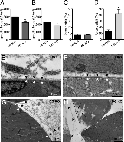
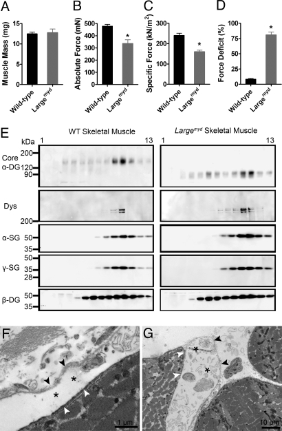
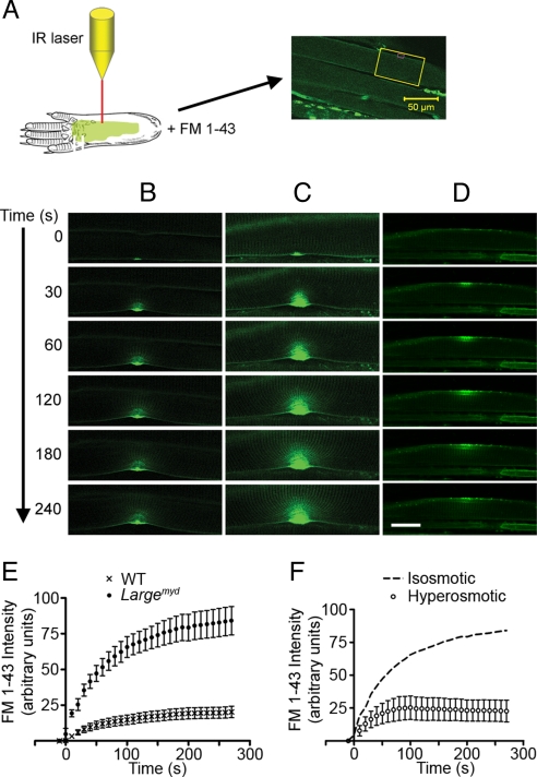
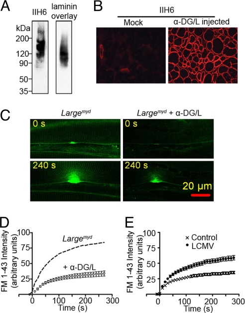
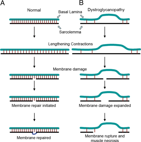
Skeletal muscle basal lamina is linked to the sarcolemma through transmembrane receptors, including integrins and dystroglycan. The function of dystroglycan relies critically on posttranslational glycosylation, a common target shared by a genetically heterogeneous group of muscular dystrophies characterized by alpha-dystroglycan hypoglycosylation. Here we show that both dystroglycan and integrin alpha7 contribute to force-production of muscles, but that only disruption of dystroglycan causes detachment of the basal lamina from the sarcolemma and renders muscle prone to contraction-induced injury. These phenotypes of dystroglycan-null muscles are recapitulated by Large(myd) muscles, which have an intact dystrophin-glycoprotein complex and lack only the laminin globular domain-binding motif on alpha-dystroglycan. Compromised sarcolemmal integrity is directly shown in Large(myd) muscles and similarly in normal muscles when arenaviruses compete with matrix proteins for binding alpha-dystroglycan. These data provide direct mechanistic insight into how the dystroglycan-linked basal lamina contributes to the maintenance of sarcolemmal integrity and protects muscles from damage.


Conflict of interest statement

The authors declare no conflict of interest.

Figures

Fig. 1.
Fig. 1.
Contractile and ultrastructural properties of DG- and α7-deficient skeletal muscle. (A–D) Specific force (A and B) and force deficit (C and D) after 2 lengthening contractions of the EDL muscle from α7-null (α7 KO) and DG-null (DG KO) mice were compared to those for littermate controls. (*, P < 0.05.) All data are presented as the mean ± SD. (E–G) Ultrastructure of quadriceps muscle from 5-week-old control (E), integrin α7-null (F), and DG-null (G) mice in the absence of exercise. (H) Ultrastructure of exercised quadriceps muscle from DG-null mice immediately after downhill treadmill running. Black arrowheads, basal lamina; white arrowhead, sarcolemma; black asterisk, site of separation of the sarcolemma and the basal lamina; dashed line, outline of the disrupted sarcolemma; black arrow, disruption of sarcomere structure.
Fig. 2.
Fig. 2.
Severe muscular dystrophy in DG/α7 DKO mice. (A) Total distance that the mice traveled within 12 h in open field activity cages. (B) Vertical movement activity. Vertical movement activity was represented as the number of rearing movements. DKO significantly impaired vertical movement compared with littermates (P < 0.01). The values in all data are averages from 3–7 mice of each group: WT (n = 7), DG-null (n = 6), α7-null (n = 4), and DKO (n = 3). (C–F) H&E staining of quadriceps sections. Severe pathological changes are observed in DKO section, including variations in fiber size, centrally located nuclei, and infiltration of inflammatory cells. White triangles, centrally nucleated cells. (G) Central nucleation is represented as the percentage of total nucleated fibers with centrally located nuclei. (H and I) Separation of the basal lamina from the sarcolemma (H) and loss of the basal lamina structure (I) in quadriceps muscles from DKO observed under electron microscopy. White arrowhead, sarcolemma; asterisk, separation of the basal lamina and the sarcolemma; black arrow, detached basal lamina; white arrow, disrupted basal lamina.
Fig. 3.
Fig. 3.
Characterization of the contractile properties and the DGC structure in the Largemyd muscle. (A–C) EDL muscle mass (A), maximum force (B), and specific force (C) before subjection to the lengthening-contraction protocol. (D) Force deficits following the lengthening-contraction protocol, as measured for EDL muscles in vitro from C57BL/6 (n = 6) and Largemyd (n = 6) mice. (*, P < 0.05.) All data are presented as the mean ± SEM. (E) Solubilized C57BL/6 and Largemyd skeletal muscle were enriched for DGC by wheat germ agglutinin (WGA) affinity chromatography and separated on 10–30% sucrose gradients. Gradient fractions (1, top; 13, bottom) were blotted with antibodies against core α-DG, dystrophin (Dys), α-sarcoglycan (SG), γ-SG, and β-DG. (F and G) Ultrastructural analysis of quadriceps muscles from Largemyd mice were observed under electron microscopy. Black arrowhead, basal lamina; white arrowhead, sarcolemma; asterisk, dissociation of basal lamina and sarcolemma.
Fig. 4.
Fig. 4.
Membrane damage assay on WT and Largemyd skeletal muscle. (A) Schematics of the in situ membrane damage assay. (B–D) Representative examples of time-lapsed images of membrane damage assay performed on C57BL/6 (B) and Largemyd skeletal muscle fibers in regular Tyrode buffer (C), or in a hyperosmotic buffer (D). (Scale bar: 20 μm.) (E) Plot of FM 1-43 fluorescence intensity against time in WT (n = 7) and Largemyd (n = 8) muscle fibers. (F) Plot of FM 1-43 fluorescence intensity against time in Largemyd (n = 5) muscle fibers in the hyperosmotic buffer. Dashed curve represents membrane damage data in Largemyd muscle in regular Tyrode buffer (isosmotic), from the experiment whose results are depicted in E. All data are presented as mean ± SEM.
Fig. 5.
Fig. 5.
Effect of α-DG-mediated association of the basal lamina with the sarcolemma on membrane integrity. (A) The purified recombinant α-DG reacted with the glycosylated α-DG antibody IIH6 (Left) and bound to laminin in the laminin overlay assay (Right). (B) The Largemyd muscles injected with recombinant α-DG/L (α-DG/L injected) or saline (Mock) were stained with IIH6 antibody. (C) Representative micrographs of membrane damage assay performed on Largemyd muscle fibers treated with or without recombinant α-DG/L. (D) Plot of FM 1-43 fluorescence intensity against time of the in situ membrane damage assay in Largemyd muscle fibers treated with recombinant α-DG/L (n = 7). The dash curve represents mean FM 1-43 fluorescence intensity of the membrane damage assay in Largemyd muscle from the Fig. 4E. (E) Plot of FM 1-43 fluorescence intensity against time for the in situ membrane damage assay carried out in C57BL/6 muscle fibers treated with (n = 9) or without (n = 11) LCMV. All of the data are means ± SEM.
Fig. 6.
Fig. 6.
A proposed mechanism for the basal-lamina-mediated prevention of membrane damage during lengthening contractions. (A) In normal skeletal muscle, the sarcolemma is tightly associated with the basal lamina. Lengthening contractions cause an increase in tension within the sarcolemma, which can lead to small membrane tears. The dysferlin-mediated membrane repair mechanism subsequently reseals the membrane and maintains membrane integrity. (B) In DG-null skeletal muscle, the tight association of the sarcolemma with the basal lamina is lost, and thus membrane tears that developed during lengthening contractions rapidly expand, leading to the loss of a large segment of the sarcolemma.

Similar articles

Cited by

References

    1. Davies KE, Nowak KJ. Molecular mechanisms of muscular dystrophies: Old and new players. Nat Rev Mol Cell Biol. 2006;7:762–773. - PubMed
    1. Durbeej M, Campbell KP. Muscular dystrophies involving the dystrophin-glycoprotein complex: An overview of current mouse models. Curr Opin Genet Dev. 2002;12:349–361. - PubMed
    1. Meryon E. On granular and fatty degeneration of voluntary muscles. Med Chir Trans. 1852;35:73–84. - PMC - PubMed
    1. Sanes JR. The basement membrane/basal lamina of skeletal muscle. J Biol Chem. 2003;278:12601–12604. - PubMed
    1. Candiello J, et al. Biomechanical properties of native basement membranes. FEBS J. 2007;274:2897–2908. - PubMed

Publication types