. 2009 Aug 11;106(32):13529-34.
doi: 10.1073/pnas.0905825106. Epub 2009 Jul 24.

The kuru infectious agent is a unique geographic isolate distinct from Creutzfeldt-Jakob disease and scrapie agents

Affiliations

The kuru infectious agent is a unique geographic isolate distinct from Creutzfeldt-Jakob disease and scrapie agents

Laura Manuelidis et al. Proc Natl Acad Sci U S A. .

Abstract

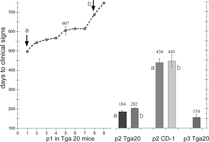
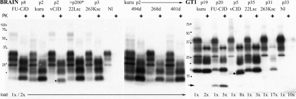
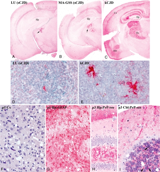
Human sporadic Creutzfeldt-Jakob disease (sCJD), endemic sheep scrapie, and epidemic bovine spongiform encephalopathy (BSE) are caused by a related group of infectious agents. The new U.K. BSE agent spread to many species, including humans, and clarifying the origin, specificity, virulence, and diversity of these agents is critical, particularly because infected humans do not develop disease for many years. As with viruses, transmissible spongiform encephalopathy (TSE) agents can adapt to new species and become more virulent yet maintain fundamentally unique and stable identities. To make agent differences manifest, one must keep the host genotype constant. Many TSE agents have revealed their independent identities in normal mice. We transmitted primate kuru, a TSE once epidemic in New Guinea, to mice expressing normal and approximately 8-fold higher levels of murine prion protein (PrP). High levels of murine PrP did not prevent infection but instead shortened incubation time, as would be expected for a viral receptor. Sporadic CJD and BSE agents and representative scrapie agents were clearly different from kuru in incubation time, brain neuropathology, and lymphoreticular involvement. Many TSE agents can infect monotypic cultured GT1 cells, and unlike sporadic CJD isolates, kuru rapidly and stably infected these cells. The geographic independence of the kuru agent provides additional reasons to explore causal environmental pathogens in these infectious neurodegenerative diseases.


Conflict of interest statement

The authors declare no conflict of interest.

Figures

Fig. 1.
Fig. 1.
Transmission of kuru Creutzfeldt–Jakob disease (kCJD) to mice. Graph shows days of incubation in the 9 intracerebrally inoculated Tga20 mice at passage 1 (p1) [607 ± 26 (SEM)]. Brain homogenates from passage 1 mice a and b with an ≈200-day difference in incubation time then were inoculated into both Tga20 and CD-1 mice. Brains a and b yield the same incubation time. Reductions in passage 2 (p2) incubations indicate agent adaptation in both mouse genotypes, with Tga20 mice showing a greater reduction. By passage 3 (p3), Tga20 mice provide a rapid kCJD model.
Fig. 2.
Fig. 2.
The prion protein (PrP) and PrP-res bands [proteinase K (PK)+ lanes] in CD-1 mouse brain homogenates and in GT1 cultures assayed on Western blots (SI Methods). Only the variant Creutzfeldt–Jakob disease (vCJD) agent shows a unique ≈19-kDa band (at dot). Kuru samples show one mouse with a long (55-day) clinical course (494-day lane) with more PrP-res than mice with typical 15–20 day signs; all kuru (kCJD) mice (268- and 401-day lanes), including Tga20 mice, show the same PrP-res profile. The 22L-sc CD-1 mouse (asterisk) was inoculated with 22L-sc-infected N2a cells displaying an N2a-specific but not agent-specific PrP band pattern (–11), yet these cells reproduced their original 22L-sc incubation time and neuropathology (–11) and the type I brain PrP pattern in mice (22L-sc lane). Protein loads and mouse passages are noted. The GT1 lanes show GT1-cell-specific PrP and PrP-res patterns (e.g., the heavily glycosylated top band in undigested brain homogenates is at 34 kDa, whereas it is at 39 kDa in GT1 homogenates). Kuru brain homogenates rapidly, reproducibly, and stably infected GT1 cells, as shown by the high levels of PrP-res (kCJD lane, >4 experiments), whereas sCJD gave only subliminal infection with the same homogenate exposure (SI Methods). The 13-kDa PrP-res marker in GT1 cells (arrow) is diagnostic for Japanese isolates in GT1 cells but not brain (FU-CJD lanes), but the 19-kDa doublet, found only in vCJD brain infections (at dot), is again seen in GT1 cultures. The band profile in kCJD is comparable with those of 22L-sc and 263K-sc in GT1 cells but required much higher amounts of protein to be loaded to each lane, as noted. Uninfected lanes (Nl) show no PrP-res.
Fig. 3.
Fig. 3.
Sporadic Creutzfeldt–Jakob disease (sCJD) and kuru (kCJD) tissue differences. The first row (A–C) shows low-magnification images of brain from passage 2 CD-1 mice with sCJD versus those with kCJD. All mice became sick at similar times (377, 384, and 403 days) yet still show that specific neuronal nuclei are affected. Both sCJD isolates (LU and MA) induce pathological prion protein (PrP) in the medial thalamus (dark red at arrow) like SY-CJD (12). Kuru infection (C) provokes extensive PrP deposits in a much larger thalamic region (Th) and in hippocampus (Hp), a band of cortical gray (Cx), and temporal lobe/Ammon's horn (Am). Profound differences in spleen PrP are seen with sCJD (D) versus kCJD (E) in passage 2 CD-1 mice. The third row (F–I) shows kCJD brains in Tga20 mice with spongiform lesions in temporal cortex at passage 1 (F), intense gliosis with red-stained astrocytes (G), and abnormal PrP deposits in hippocampus at passage 3 (H). Cerebellum (I) shows pathological PrP deposits consistent with degenerating synaptic boutons (arrows) that become more prominent with further digestion (SI Methods) (i.e., they contain PrP amyloid).

References

    1. Gajdusek DC. Unconventional viruses and the origin and disappearance of kuru. Science. 1977;197:943–960. - PubMed
    1. Hadlow WJ. Scrapie and kuru. Lancet. 1959;274:289–290.
    1. Sigurdsson B. Rida: A chronic encephalitis of sheep. With general remarks on infections which develop slowly and some of their characteristics. Br Vet J. 1954;110:341–354.
    1. Hunter N, Cairns D. Scrapie-free Merino and Poll Dorset sheep from Australia and New Zealand have normal frequencies of scrapie-susceptible PrP genotypes. J Gen Virol. 1998;79:2079–2082. - PubMed
    1. Manuelidis E, Kim J, Angelo J, Manuelidis L. Serial propagation of Creutzfeldt–Jakob disease in guinea pigs. Proc Natl Acad Sci USA. 1976;73:223–227. - PMC - PubMed

Publication types