. 2009 Aug 11;106(32):13335-40.
doi: 10.1073/pnas.0812911106. Epub 2009 Jul 24.

NFAT isoforms control activity-dependent muscle fiber type specification

Affiliations

NFAT isoforms control activity-dependent muscle fiber type specification

Elisa Calabria et al. Proc Natl Acad Sci U S A. .

Abstract

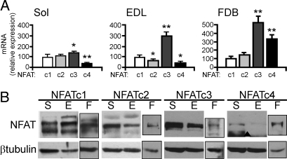
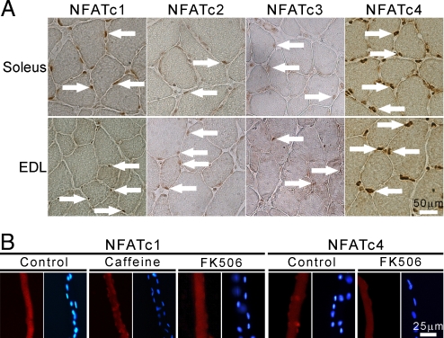
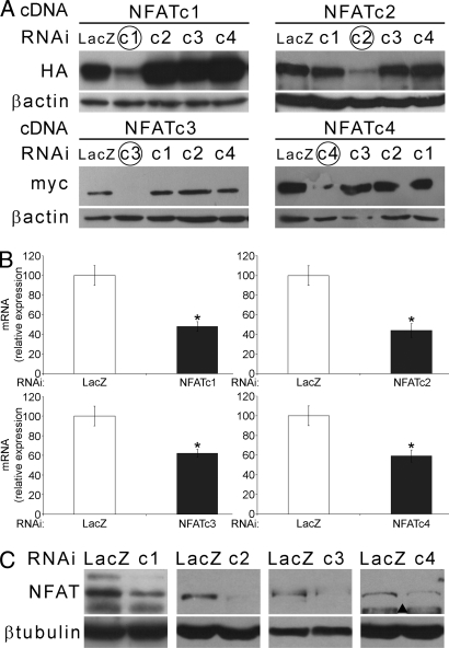
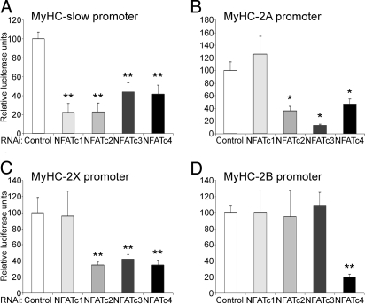
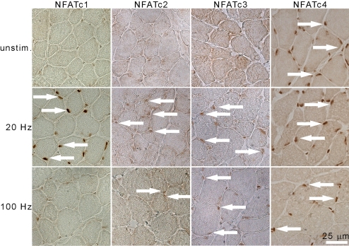
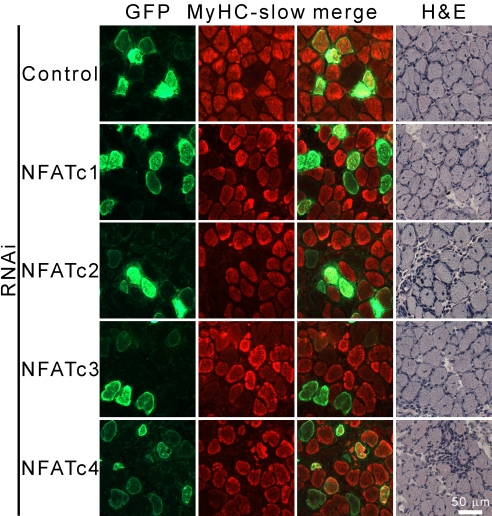
The intracellular signals that convert fast and slow motor neuron activity into muscle fiber type specific transcriptional programs have only been partially defined. The calcium/calmodulin-dependent phosphatase calcineurin (Cn) has been shown to mediate the transcriptional effects of motor neuron activity, but precisely how 4 distinct muscle fiber types are composed and maintained in response to activity is largely unknown. Here, we show that 4 nuclear factor of activated T cell (NFAT) family members act coordinately downstream of Cn in the specification of muscle fiber types. We analyzed the role of NFAT family members in vivo by transient transfection in skeletal muscle using a loss-of-function approach by RNAi. Our results show that, depending on the applied activity pattern, different combinations of NFAT family members translocate to the nucleus contributing to the transcription of fiber type specific genes. We provide evidence that the transcription of slow and fast myosin heavy chain (MyHC) genes uses different combinations of NFAT family members, ranging from MyHC-slow, which uses all 4 NFAT isoforms, to MyHC-2B, which only uses NFATc4. Our data contribute to the elucidation of the mechanisms whereby activity can modulate the phenotype and performance of skeletal muscle.


Conflict of interest statement

The authors declare no conflict of interest.

Figures

Fig. 1.
Fig. 1.
Expression and localization of NFAT isoforms in skeletal muscle. (A) Quantitative PCR of Soleus (Sol), EDL, and isolated fibers from FDB. Results are expressed as percentage of NFATc1 expression. (B) WB with antibodies specific for NFAT isoforms of total muscle extracts from Soleus (S), EDL (E), and FDB fibers (F). ▴, aspecific band.
Fig. 2.
Fig. 2.
Localization of NFAT isoforms in adult muscle. (A) Cross-sections of soleus and EDL stained with antibodies specific for all 4 NFAT isoforms as indicated. White arrows indicate some positive nuclei. (B) Cultured muscle fibers from FDB stained with anti-NFATc1 and anti-NFATc4 Abs, and corresponding counterstaining of nuclei with Hoechst; treatments, FK506 (1 μM), caffeine (10 mM), 2 h.
Fig. 3.
Fig. 3.
RNAi of individual NFATs. (A) WB showing specific down-regulation of the target NFAT (HA or myc-tagged as indicated), but not of other isoforms obtained by cotransfection of NFAT cDNA and isoform-specific siRNAs in HEK293 cells. Down-regulation of endogenous NFATs in muscles transfected with NFAT isoform-specific and control siRNAs is shown by qPCR at the RNA (B) and by WB at the protein level (C). ▴, aspecific band.
Fig. 4.
Fig. 4.
Role of individual NFATs in fiber type specific MyHC expression. The promoters of the MyHC isoforms linked to luciferase were cotransfected with siRNAs specific for either GFP or LacZ (control) or for the indicated NFAT isoforms. All experiments are means of at least 4 individual muscles per data point, repeated with 2 siRNA sequences.
Fig. 5.
Fig. 5.
Nucleocytoplasmic shuttling of NFAT isoforms in response to electrical activity. IHC staining of endogenous NFAT isoforms in cross-sections of EDL electrically stimulated with patterns of impulses at 20 and 100 Hz as indicated, for 2 h. Contralateral EDL from the anesthetized animal was used as unstimulated control (unstim). White arrows indicate NFAT-positive nuclei.
Fig. 6.
Fig. 6.
All NFAT isoforms are necessary for the expression of MyHC-slow in regenerating muscle. Regenerating soleus was cotransfected with NFAT isoform-specific siRNAs and SNAP-GFP at day 3 after myotoxic damage induced with bupivacaine. Analyses were performed 1 week later. Serial cross-sections were stained with antibodies specific for MyHC-slow and GFP or with hematoxylin/eosin (H&E).

References

    1. Buckingham M, et al. The formation of skeletal muscle: from somite to limb. J Anat. 2003;202:59–68. - PMC - PubMed
    1. Schiaffino S, Sandri M, Murgia M. Activity-dependent signaling pathways controlling muscle diversity and plasticity. Physiology. 2007;22:269–278. - PubMed
    1. Hughes SM, et al. Selective accumulation of MyoD and myogenin mRNAs in fast and slow adult skeletal muscle is controlled by innervation and hormones. Development. 1993;118:1137–1147. - PubMed
    1. Wheeler MT, Snyder EC, Patterson MN, Swoap SJ. An E-box within the MHC IIB gene is bound by MyoD and is required for gene expression in fast muscle. Am J Physiol. 1999;276:C1069–C1078. - PubMed
    1. Ekmark M, Rana ZA, Stewart G, Hardie DG, Gundersen K. De-phosphorylation of MyoD is linking nerve-evoked activity to fast myosin heavy chain expression in rodent adult skeletal muscle. J Physiol. 2007;584:637–650. - PMC - PubMed

Publication types

LinkOut - more resources