. 2009 Aug 11;106(32):13594-9.
doi: 10.1073/pnas.0901402106. Epub 2009 Jul 24.

Neural stem cells improve cognition via BDNF in a transgenic model of Alzheimer disease

Affiliations

Neural stem cells improve cognition via BDNF in a transgenic model of Alzheimer disease

Mathew Blurton-Jones et al. Proc Natl Acad Sci U S A. .

Abstract

Neural stem cell (NSC) transplantation represents an unexplored approach for treating neurodegenerative disorders associated with cognitive decline such as Alzheimer disease (AD). Here, we used aged triple transgenic mice (3xTg-AD) that express pathogenic forms of amyloid precursor protein, presenilin, and tau to investigate the effect of neural stem cell transplantation on AD-related neuropathology and cognitive dysfunction. Interestingly, despite widespread and established Ass plaque and neurofibrillary tangle pathology, hippocampal neural stem cell transplantation rescues the spatial learning and memory deficits in aged 3xTg-AD mice. Remarkably, cognitive function is improved without altering Ass or tau pathology. Instead, the mechanism underlying the improved cognition involves a robust enhancement of hippocampal synaptic density, mediated by brain-derived neurotrophic factor (BDNF). Gain-of-function studies show that recombinant BDNF mimics the beneficial effects of NSC transplantation. Furthermore, loss-of-function studies show that depletion of NSC-derived BDNF fails to improve cognition or restore hippocampal synaptic density. Taken together, our findings demonstrate that neural stem cells can ameliorate complex behavioral deficits associated with widespread Alzheimer disease pathology via BDNF.


Conflict of interest statement

The authors declare no conflict of interest.

Figures
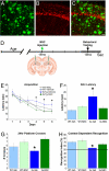
Fig. 1.
Fig. 1.
Neural stem cell transplantation improves AD-related cognitive dysfunction. Eighteen-month-old 3xTg-AD mice exhibit robust plaques (A and C; green) and tangles (B; red) within the hippocampus. (D) 100,000 GFP-NSCs or vehicle control were stereotactically injected. Four weeks later, learning and memory was tested. (E) MWM training revealed that all groups learn the task. However, NSC-injected 3xTg-AD mice exhibit significantly shorter escape latencies on days 4–6 of training vs. vehicle-injected transgenics (ANOVA, P < 0.04, FPLSD P < 0.029). (F) In probe trial testing, NSC-injected 3xTg-AD mice also achieve significantly shorter latencies than vehicle-injected 3xTg-AD mice and perform equivalent to nonTg controls (ANOVA, P = 0.042, FPLSD P = 0.010). (G) Likewise, NSC-injected 3xTg-AD mice cross the former platform location more often than control-injected transgenics (ANOVA, P = 0.014, FPLSD P = 0.002). (H) Context-dependent object recognition testing reveals that vehicle-injected 3xTg-AD mice are impaired, spending an equivalent amount of time exploring both objects. In contrast, NSC-injected 3xTg-AD mice exhibit a partial but significant recovery in this task (ANOVA, P = 0.0047, FPLSD P = 0.041, vs. vehicle-injected 3xTg-AD mice). Data presented as mean ± SEM. (Scale bar, 45 μm.)
Fig. 2.
Fig. 2.
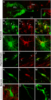
Engrafted neural stem cells differentiate into neurons, astrocytes, and oligodendrocytes. (A) GFP-expressing NSCs (green) migrated from their hippocampal injection site and engrafted predominately within 2 major regions, either surrounding the granule cell layer (GCL) of the dentate gyrus or within white matter tracts including the fimbria fornix and corpus callosum. Confocal microscopy revealed only limited chemotaxis of NSCs toward Aβ plaques (red; A Inset). LV: lateral ventricle, Sub: subiculum. Engrafted NSCs differentiated into all 3 lineages. (B–G) The majority of NSCs (39.4%) differentiated into astrocytes, coexpressing GFAP (red). (H–J) Within white matter tracts, NSCs often exhibited oligodendroglial morphology and 26.4% coexpressed the oligodendroglial marker (GalC; red). (K–M) Far fewer NSCs adopted a neuronal fate (5.8%) as evidenced by expression of doublecortin (red). (N) neuronally-differentiated NSCs with pyramidal cell morphology surrounded by presynaptic terminals (synaptophysin, red), or exhibiting dendritic spine architecture (O–P) were also occasionally observed. (Scale bars, 80 μm in A, 12 μm in B–D, 5 μm in E–G and N, 15 μm in H–J, 7 μm in K–M, 1.5 μm in O and 200 nm in P.)
Fig. 3.
Fig. 3.
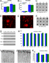
Aβ and tau pathology is not altered by neural stem cell transplantation. Biochemical and histological analyses reveal no differences between vehicle- and NSC-injected 3xTg-AD mice in Aβ and tau pathology. ELISA for soluble (A) and insoluble (B) levels of Aβ40 and Aβ42 reveal no differences in brain Aβ load (ANOVA P = 0.69, mean ± SEM). (C) Likewise, dot-blot analysis with the oligomer-specific antibody A11 revealed no changes in soluble oligomers (P = 0.36). (D–E) Immunofluorescent microscopy demonstrated no differences in plaque load within the hippocampus of vehicle-injected (D) versus NSC-injected (E) mice, quantified in (H) (P = 0.52). (F) Dot-blot analysis with the fibril-specific antibody OC also revealed no changes (P = 0.40). (G). Immunoblot analysis of APP, IDE, total human tau, and PHF-1 phosphorylated tau (S396/S404) also revealed no differences between groups (P > 0.56). Quantification in (H) is normalized to control levels ± SEM. (G–I) Immunofluorescent assessment of phosphorylated tau (S199/S202) within CA1 pyramidal neurons also revealed no differences in the somatodendritic localization of tau between vehicle- (I) and NSC- (J) injected mice; quantification in (K). Vehicle and NSC-injected samples denoted by V and N respectively. Pyr: CA1 pyramidal cell layer, SR: stratum radiatum. (Scale bars, 30 μm in D–E, 25 μm in I–J.)
Fig. 4.
Fig. 4.
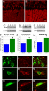
Neural stem cells increase synaptic density and produce BDNF. (A–C) Confocal optical densitometry revealed that compared to vehicle-injected 3xTg-AD mice (A), NSC-injected 3xTg-AD mice (B) exhibit a 67% increase in synaptophysin immunoreactivity within the stratum radiatum of CA1 (red puncta, quantified in F, P = 0.0028). (C) Immunoblot analysis of vehicle-injected (C) and NSC-injected 3xTg-AD mice (N) also revealed a significant 45% increase in synaptophysin following NSC transplantation (quantified in G, P = 0.01). (D) Comparison of NSC-derived (N) and control neuronal cell line (C) cultures revealed a high level of BDNF expression within NSCs. (E) In vivo analysis by Western blot and further quantification by ELISA (H) demonstrated a significant elevation of BDNF in NSC-injected mice (N) versus control-injected mice (C) (P = 0.047). (I) Confocal microscopy for GFP-NSCs (F) and pro-BDNF (J) demonstrate that engrafted NSCs often coexpress pro-BDNF (K). Expression of pro-BDNF within adjacent non-GFP-expressing endogenous cells was also observed (K, red only). (L–Q) Colocalization between GFP NSCs (green) and total BDNF was also observed. Data in F–H is normalized to control levels and shown as mean ± SEM. (Scale bars, 10 μm in A and B, 18 μm in I–K, 3 μm in L–Q.)
Fig. 5.
Fig. 5.
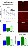
BDNF is necessary for NSC-induced cognitive rescue and increased synaptic density. (A) NSC BDNF expression was reduced 78% by shRNA knockdown (P = 0.0006). Eighteen-month-old 3xTg-AD mice were injected with BDNF-shRNA NSCs or control NSCs and 1 month later tested in the MWM. (B) Control NSCs improved cognition leading to shorter escape latencies on days 3–6 of training (ANOVA P = 0.027, FPLSD P < 0.03). In contrast, BDNF-shRNA NSC-injected mice showed no improvement and were impaired vs. control NSC-injected animals (FPLSD P < 0.013). In probe trial testing (C) control NSC-injected animals found the former platform location significantly faster than vehicle-injected mice (FPLSD P = 0.037), whereas BDNF-shRNA NSC-injected mice performed at an intermediate level that was not significantly different from vehicle-injected mice (FPLSD P = 0.29). (D) Likewise, control NSC-injected mice crossed the former platform location significantly more than the other 2 groups (ANOVA P = 0.0049, FPLSD P < 0.0136). To determine whether NSC-derived BDNF also mediates the observed changes in synaptic density, synaptophysin was quantified. Vehicle-injected animals (E) exhibited significantly less immunoreactive puncta than control NSC-injected animals (F, ANOVA, P < 0.0001, FPLSD P = 0.0001). In contrast, BDNF-shRNA NSC-injected animals exhibited an intermediate level of synaptic density (G), significantly greater than vehicle-injected mice (P = 0.014) but also less than control NSC-injected mice (P = 0.0093). (H) Significance vs. vehicle-injected mice denoted by *, vs. BDNF-shRNA NSC-injected mice denoted by #. (Scale bar, 10 μm.)

References

    1. Ebert AD, Beres AJ, Barber AE, Svendsen CN. Human neural progenitor cells over-expressing IGF-1 protect dopamine neurons and restore function in a rat model of Parkinson's disease. Exp Neurol. 2008;209:213–223. - PubMed
    1. Lu P, Jones LL, Snyder EY, Tuszynski MH. Neural stem cells constitutively secrete neurotrophic factors and promote extensive host axonal growth after spinal cord injury. Exp Neurol. 2003;181(2):115–129. - PubMed
    1. Park KI, et al. Neural stem cells may be uniquely suited for combined gene therapy and cell replacement: Evidence from engraftment of Neurotrophin-3-expressing stem cells in hypoxic-ischemic brain injury. Exp Neurol. 2006;199(1):179–190. - PubMed
    1. Billings LM, Oddo S, Green KN, McGaugh JL, LaFerla FM. Intraneuronal Abeta causes the onset of early Alzheimer's disease-related cognitive deficits in transgenic mice. Neuron. 2005;45(5):675–688. - PubMed
    1. Oddo S, et al. Triple-transgenic model of Alzheimer's disease with plaques and tangles: Intracellular Abeta and synaptic dysfunction. Neuron. 2003;39(3):409–421. - PubMed

Publication types

MeSH terms

Substances