. 2009 Sep 17;114(12):2401-10.
doi: 10.1182/blood-2009-04-214619. Epub 2009 Jul 24.

Targeted disruption of Zfp36l2, encoding a CCCH tandem zinc finger RNA-binding protein, results in defective hematopoiesis

Affiliations

Targeted disruption of Zfp36l2, encoding a CCCH tandem zinc finger RNA-binding protein, results in defective hematopoiesis

Deborah J Stumpo et al. Blood. .

Abstract

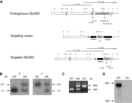
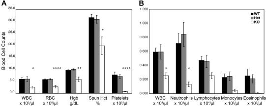
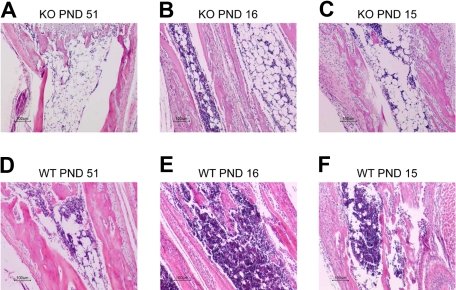
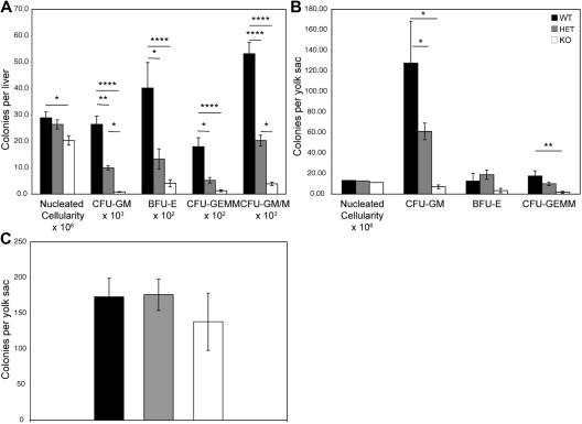
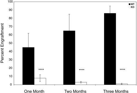
Members of the tristetraprolin family of tandem CCCH finger proteins can bind to AU-rich elements in the 3'-untranslated region of mRNAs, leading to their deadenylation and subsequent degradation. Partial deficiency of 1 of the 4 mouse tristetraprolin family members, Zfp36l2, resulted in complete female infertility because of early embryo death. We have now generated mice completely deficient in the ZFP36L2 protein. Homozygous Zfp36l2 knockout (KO) mice died within approximately 2 weeks of birth, apparently from intestinal or other hemorrhage. Analysis of peripheral blood from KO mice showed a decrease in red and white cells, hemoglobin, hematocrit, and platelets. Yolk sacs from embryonic day 11.5 (E11.5) Zfp36l2 KO mice and fetal livers from E14.5 KO mice gave rise to markedly reduced numbers of definitive multilineage and lineage-committed hematopoietic progenitors. Competitive reconstitution experiments demonstrated that Zfp36l2 KO fetal liver hematopoietic stem cells were unable to adequately reconstitute the hematopoietic system of lethally irradiated recipients. These data establish Zfp36l2 as a critical modulator of definitive hematopoiesis and suggest a novel regulatory pathway involving control of mRNA stability in the life cycle of hematopoietic stem and progenitor cells.


Figures

Figure 1
Figure 1
Targeted disruption of Zfp36l2. (A) Schematic representation of the normal genomic locus for Zfp36l2 as well as the targeting vector and resulting disrupted allele generated by homologous recombination. The 2 exons are represented by gray boxes. The translational start site is indicated by the arrow (under Sa). Genomic 5′ and 3′ probes, located outside the targeting vector, are represented by stippled boxes. The targeting vector contained a neomycin resistance marker cassette (PGKNeo), causing the deletion of a large portion of the second exon. A diphtheria toxin resistance element was also inserted into the targeting vector (PGKDTA). The black lines above the endogenous and targeted genes represent the expected fragment length after digestion with EcoRV and SstI or EcoRI and hybridization with the 5′ probe or 3′ probe, respectively. PCR primers used to detect homologous recombination (F1, R1) as well as for genotyping offspring (F1, F2, and R2) are indicated by the arrows beneath the endogenous and targeted genes. Primers F3, R3, F4, and R4 were used to generate an exon one fragment (F3, R3) or a 3′UTR fragment (F4, R4) used as hybridization probes for Northern blots. Abbreviations for restriction enzyme sites are as follows: A indicates Asp718; C, Csp45I; E, EcoRI; EV, EcoRV; H, HindIII; N, NotI; Sa, SalI; S, SstI; St; stop codon; X, XbaI. (B) Southern blot analysis of EcoRV/SstI (5′ genomic probe) and EcoRI (3′ genomic probe)–digested genomic DNA from WT and homologously recombined (HR) ES cells. (C) PCR analysis of genomic DNA isolated from Zfp36l2 WT, heterozygous, and KO mice using PCR primers F1, F2, and R2. (D) Northern blot of total cellular RNA (15 μg) isolated from E14.5 WT and KO fetal liver and hybridized with a 32P-labeled 787-bp 3′UTR probe for Zfp36l2.
Figure 2
Figure 2
Developmental expression and tissue distribution of Zfp36l2 mRNA. Total cellular RNA (15 μg) was isolated from yolk sacs, placentas, and whole embryos at the indicated gestational stages (A) and from adult mouse tissues and E14.5 liver (B). Northern blots were hybridized with a 32P-labeled 787-bp 3′UTR probe for Zfp36l2. The positions of Zfp36l2 mRNA and the ribosomal RNAs are shown to the left of the blot. The bottom panels show acridine orange staining of the samples as loading controls.
Figure 3
Figure 3
In situ hybridization histochemistry of Zfp36l2 mRNA expression at E14.5. Sagittal sections of E14.5 WT and KO embryos were hybridized with Zfp36l2 (A) or GAPDH (B) antisense probes. The specificity of the Zfp36l2 antisense probe was demonstrated by the absence of signal seen with the KO embryo compared with the WT embryo. A sense probe for Zfp36l2 produced essentially no signal at this exposure time (data not shown). Note the weak hybridization seen in the KO placenta in the maternal decidua, a tissue that should be heterozygous for Zfp36l2. (C) Neighboring sections stained with hematoxylin and eosin. b indicates brain; d, decidua; h, heart; l, liver; ys, yolk sac; and p, placenta. Original magnifications ×40, obtained using the Aperio Scanscope T2 Scanner (Aperio Technologies Inc). Scanned images were imported into Aperio Imagescope, Version 6.25.0.1117.
Figure 4
Figure 4
Hematologic analysis of WT, heterozygous, and KO mice. (A) Complete blood cell counts of peripheral blood from Zfp36l2 WT (n = 6), heterozygous (n = 7), and KO (n = 6) mice at 14 to 15 days of age. (B) White blood cell differential counts for peripheral blood from Zfp36l2 WT (n = 6), heterozygous (n = 7), and KO (n = 5, except WBC, n = 6) mice. Values are mean ± SEM. Statistical significance was determined using one-way analysis of variance with Tukey HSD post hoc test: *P < .05; **P < .01; ****P < .001. WBC indicates white blood cell; RBC, red blood cell; Hgb, hemoglobin; and Hct, hematocrit.
Figure 5
Figure 5
Histologic analysis of bone marrow in Zfp36l2 KO and WT mice. Tissue sections from the radius/ulna of PND 51 KO (A) and WT (D) mice, PND 16 KO (B) and WT (E) mice, and PND 15 KO (C) and WT (F) littermate pairs of mice were stained with hematoxylin and eosin. Note the hypocellular bone marrow along with an increased number of adipocytes in the KO samples. Original magnifications ×10, obtained with a Nikon Eclipse E600 microscope with a Nikon DXM1200 digital camera. Images were imported into the Nikon Act-1 software.
Figure 6
Figure 6
Hematopoietic progenitor colony assays from fetal liver and yolk sacs of Zfp36l2 WT, heterozygous, and KO embryos. Fetal liver cells (A) isolated from E14.5 embryos (WT, n = 15; heterozygous, n = 26, KO, n = 14) and yolk sac cells (B) isolated from E11.5 embryos (WT, n = 5; heterozygous, n = 10; KO, n = 5) were grown in methylcellulose-based medium as described in “Colony-forming cell assays.” The numbers of CFU-GM, BFU-E, and CFU-GEMM, and CFU-GM/M per fetal liver or yolk sac were determined after 7 days of culture. (C) Yolk sac cells isolated from E8 to E8.25 embryos (WT, n = 20; heterozygous, n = 31; KO, n = 10) were grown in methylcellulose-based medium. Primitive erythroid progenitor colonies, EryP-colony–forming cells (C) were determined after 5 days in culture. Values are mean ± SEM. Statistical significance was determined using one-way analysis of variance with Tukey HSD post hoc test: *P < .05; **P < .01; ****P < .001.
Figure 7
Figure 7
Competitive hematopoietic reconstitution with Zfp36l2 WT and KO fetal liver cells. A total of 5 × 10⁵ donor-derived fetal liver cells (CD45.2+) from WT (n = 19) and KO (n = 23) E14.5 embryos were mixed with 5 × 10⁵ adult bone marrow recipient–derived cells from B6.BoyJ (CD45.1+) mice and were injected into lethally irradiated B6.BoyJ (CD45.1+) mice. The percentage of donor CD45.2+ cells that engrafted, as determined by CD45.1/CD45.2 staining of peripheral blood, was examined at 1, 2, and 6 months after transplantation. Values are mean ± SD. Statistical significance was determined using the Student t test: ****P < .001.

References

    1. Lai WS, Stumpo DJ, Blackshear PJ. Rapid insulin-stimulated accumulation of an mRNA encoding a proline-rich protein. J Biol Chem. 1990;265(27):16556–16563. - PubMed
    1. Carballo E, Lai WS, Blackshear PJ. Feedback inhibition of macrophage tumor necrosis factor-alpha production by tristetraprolin. Science. 1998;281(5379):1001–1005. - PubMed
    1. Taylor GA, Carballo E, Lee DM, et al. A pathogenetic role for TNF alpha in the syndrome of cachexia, arthritis, and autoimmunity resulting from tristetraprolin (TTP) deficiency. Immunity. 1996;4(5):445–454. - PubMed
    1. Carballo E, Blackshear PJ. Roles of tumor necrosis factor-alpha receptor subtypes in the pathogenesis of the tristetraprolin-deficiency syndrome. Blood. 2001;98(8):2389–2395. - PubMed
    1. Carballo E, Lai WS, Blackshear PJ. Evidence that tristetraprolin is a physiological regulator of granulocyte-macrophage colony-stimulating factor messenger RNA deadenylation and stability. Blood. 2000;95(6):1891–1899. - PubMed

Publication types

MeSH terms