

Brain anatomical abnormalities in high-risk individuals, first-episode, and chronic schizophrenia: an activation likelihood estimation meta-analysis of illness progression
- PMID: 19633214
- PMCID: PMC3004195
- DOI: 10.1093/schbul/sbp073
Brain anatomical abnormalities in high-risk individuals, first-episode, and chronic schizophrenia: an activation likelihood estimation meta-analysis of illness progression
Abstract
Objective: The present study reviewed voxel-based morphometry (VBM) studies on high-risk individuals with schizophrenia, patients experiencing their first-episode schizophrenia (FES), and those with chronic schizophrenia. We predicted that gray matter abnormalities would show progressive changes, with most extensive abnormalities in the chronic group relative to FES and least in the high-risk group.
Method: Forty-one VBM studies were reviewed. Eight high-risk studies, 14 FES studies, and 19 chronic studies were analyzed using anatomical likelihood estimation meta-analysis.
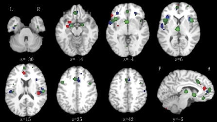
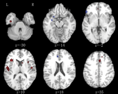
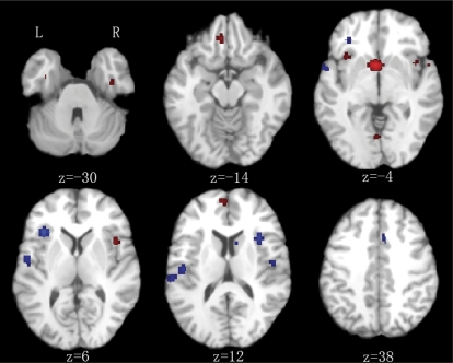
Results: Less gray matter in the high-risk group relative to controls was observed in anterior cingulate regions, left amygdala, and right insula. Lower gray matter volumes in FES compared with controls were also found in the anterior cingulate and right insula but not the amygdala. Lower gray matter volumes in the chronic group were most extensive, incorporating similar regions to those found in FES and high-risk groups but extending to superior temporal gyri, thalamus, posterior cingulate, and parahippocampal gryus. Subtraction analysis revealed less frontotemporal, striatal, and cerebellar gray matter in FES than the high-risk group; the high-risk group had less gray matter in left subcallosal gyrus, left amygdala, and left inferior frontal gyrus compared with FES. Subtraction analysis confirmed lower gray matter volumes through ventral-dorsal anterior cingulate, right insula, left amygdala and thalamus in chronic schizophrenia relative to FES.
Conclusions: Frontotemporal brain structural abnormalities are evident in nonpsychotic individuals at high risk of developing schizophrenia. The present meta-analysis indicates that these gray matter abnormalities become more extensive through first-episode and chronic illness. Thus, schizophrenia appears to be a progressive cortico-striato-thalamic loop disorder.
Figures
References
-
- Steen RG, Mull C, McClure R, Hamer RM, Lieberman JA. Brain volume in first-episode schizophrenia: systematic review and meta-analysis of magnetic resonance imaging studies. Br J Psychiatry. 2006;188:510–518. - PubMed
-
- Vita A, De Peri L, Silenzi C, Dieci M. Brain morphology in first-episode schizophrenia: a meta-analysis of quantitative magnetic resonance imaging studies. Schizophr Res. 2006;82:75–88. - PubMed
-
- Vita A, De Peri L. Hippocampal and amygdala volume reductions in first-episode schizophrenia. Br J Psychiatry. 2007;190:271. - PubMed
Publication types
MeSH terms
LinkOut - more resources
Full Text Sources
Medical
Miscellaneous
