. 2009 Sep;8(9):1449-59.
doi: 10.1128/EC.00153-09. Epub 2009 Jul 24.

Slt2 and Rim101 contribute independently to the correct assembly of the chitin ring at the budding yeast neck in Saccharomyces cerevisiae

Affiliations

Slt2 and Rim101 contribute independently to the correct assembly of the chitin ring at the budding yeast neck in Saccharomyces cerevisiae

Alberto Gomez et al. Eukaryot Cell. 2009 Sep.

Abstract

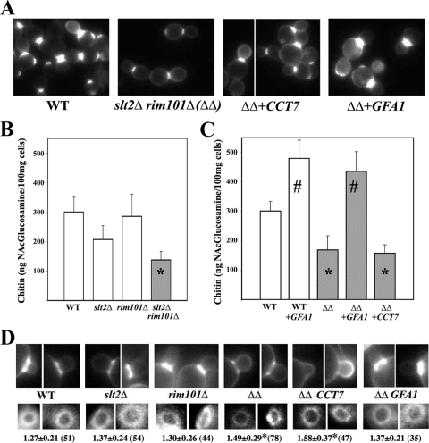
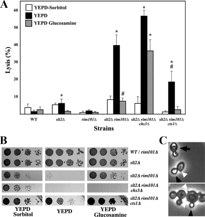
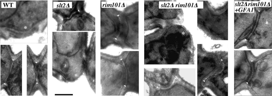
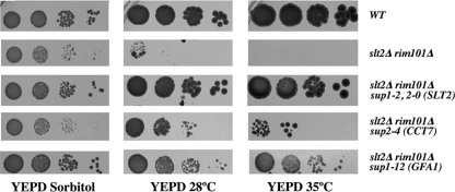
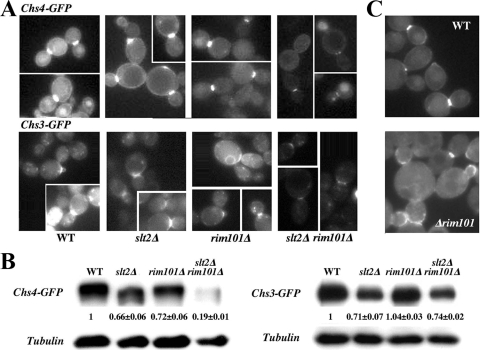
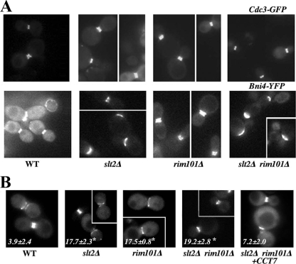
In Saccharomyces cerevisiae, the simultaneous absence of Slt2 and Rim101 prevents growth in nonosmotically stabilized media (F. Castrejon et al., Eukaryot. Cell 5:507-517, 2006). The double mutant slt2Delta rim101Delta displays altered chitin rings, together with a significant reduction in the overall levels of chitin. Cultures of this mutant lyse upon transfer to nonosmotically stabilized media, mostly through the bud, and such lysis is partially prevented by deletion of the chitinase gene (CTS1). Growth of the slt2Delta rim101Delta double mutant was restored by the overexpression of the GFA1 or CCT7 genes, which code for two biologically unrelated proteins. Further characterization of the mutant and its suppressors indicated that both Slt2 and Rim101 were independently required for the correct assembly of the septum machinery and that their concomitant absence reduced Chs3 accumulation at the neck, leading to lower levels of chitin. GFA1 overexpression, as well as the addition of glucosamine to the growth medium, specifically suppressed the growth defects by activating chitin synthesis at the neck and restoring the normal assembly of the chitin ring. In contrast, overexpression of CCT7, a Cct chaperonin subunit, alleviated the defect in the septum machinery without affecting chitin synthesis. Both suppressors thus act by reducing neck fragility through different mechanisms and allow growth in nonstabilized media. This work reports new roles for Slt2 and Rim101 in septum formation in budding yeast and confirms the homeostatic role of the chitin ring in the maintenance of neck integrity during cell division.


Figures

FIG. 1.
FIG. 1.
Chitin synthesis in selected strains. (A) Calcofluor staining of the double mutant (ΔΔ) transformed with the indicated suppressors. The wt is shown as a control. (B, C) Chitin levels in the indicated strains. Values are the means from three independent experiments, and standard deviation bars are indicated. Data marked with asterisks indicate values that are significantly (P < 0.05) different from those of the wt, and data marked with number signs indicate values that are significantly (P < 0.05) different from those of the wt or the double mutant. (D) Staining of the chitin ring after 5 min of calcofluor treatment in the indicated strains. All images were processed identically to preserve relative intensities (top row). The numbers indicate the inner diameter of the ring expressed in micrometers (bottom row). Standard deviations, as well as the number of rings measured for each strain (in parentheses), are indicated. Values that are significantly different (P < 0.05) from those of the wt are indicated with an asterisk.
FIG. 2.
FIG. 2.
Cell lysis in nonosmotically stabilized media. (A) Percentage of lysis after transferring logarithmically growing cells of the indicated strains from YEPD-sorbitol medium to the indicated media. Results were scored 3 h after the transfer, and lysis was determined by counting refringent cells using phase-contrast microscopy. The results presented are the means from two independent experiments, and standard deviations are indicated as bars. The degree of lysis observed in each medium was compared statistically with the values obtained for the wt in the corresponding media. Values significantly different (P < 0.05) from those of the corresponding wt are marked with an asterisk. A number sign indicates a significant (P < 0.05) reduction in lysis compared with the degree of lysis for the double mutant grown in YEPD. (B) Growth of the indicated mutants in different media. Logarithmically growing cells in YEPD-sorbitol were spotted onto the indicated media at a 1/10 serial dilution. Plates were incubated at 28°C for 48 h. (C) Overall aspect of the slt2Δ rim101Δ mutant grown in YEPD. Note the higher proportion of small daughter cells lysed (black arrowheads) than paired cells lysed (arrows). Note also the prominence of some bud scars in the double mutant (white arrowheads).
FIG. 3.
FIG. 3.
Ultrastructure of the yeast septum. TEM images of cells showing the septum structure in different strains. Note the more prominent scars (*) in the slt2Δ rim101Δ double mutant compared to those in the wt or slt2Δ strains and the irregular shape of the primary septum, observed as a white region between septal walls (arrowheads). Also note the more tubular aspect of the neck in the double mutant. The bar corresponds to 0.5 μm.
FIG. 4.
FIG. 4.
Suppression of the synthetic lethality of the slt2Δ rim101Δ mutant. (A) Growth of the slt2Δ rim101Δ mutant transformed with the different multicopy suppressors. Logarithmically growing cells in SC-sorbitol were spotted onto the indicated media at a 1/10 serial dilution. Plates were incubated at the indicated temperatures, and growth was scored 48 h after inoculation.
FIG. 5.
FIG. 5.
Intracellular behavior of CSIII proteins. (A) Localization of the indicated proteins in the different strains. The images of each protein were acquired under identical conditions, and pictures were processed in parallel for comparative purposes. The intensity of the label should therefore roughly reflect the relative amount of a protein in the different strains. Note the reduced intensity of both proteins in the double mutant. (B) Total protein levels, as determined by Western blotting. Numbers indicate the relative levels of Chs4-GFP or Chs3-GFP, using tubulin as a loading control. The data presented are the averages from two independent experiments. (C) Localization of Chs4-GFP integrated into the chromosome. Cells in the experiments shown in panels A and B were grown in sorbitol-supplemented SC, while the cells shown here were grown in plain SC. Note the absence of Chs4-GFP along the PM in all rim101Δ strains and the significant delocalization (see the text) of the integrated Chs4-GFP to the bud shown here.
FIG. 6.
FIG. 6.
Intracellular distribution of septum proteins. (A) Localization of the indicated proteins in the different strains. The images of each protein were acquired under identical conditions, and pictures were processed in parallel for comparative purposes. The intensity of the label should therefore roughly reflect the relative amount of a protein in the different strains. The reduced number of septin double rings in the slt2Δ and slt2Δ rim101Δ strains is appreciable in the images, but a quantitative analysis is shown in Table 1. (B) Asymmetric distribution of Cdc3 in the indicated strains. Values indicate the numbers of cells showing double rings in which either the diameter or the intensity of one of the rings is significantly different from the other, as highlighted in the images. The results are the averages from two independent experiments (n > 123), and values significantly different (P < 0.05) from those of the wt are marked with asterisks. Cells were grown in SC-sorbitol medium.

Similar articles

Cited by

References

    1. Andrews, P. D., and M. J. Stark. 2000. Type 1 protein phosphatase is required for maintenance of cell wall integrity, morphogenesis and cell cycle progression in Saccharomyces cerevisiae. J. Cell Sci. 113:507-520. - PubMed
    1. Baetz, K., J. Moffat, J. Haynes, M. Chang, and B. Andrews. 2001. Transcriptional coregulation by the cell integrity mitogen-activated protein kinase Slt2 and the cell cycle regulator Swi4. Mol. Cell. Biol. 21:6515-6528. - PMC - PubMed
    1. Baladron, V., S. Ufano, E. Duenas, A. B. Martin-Cuadrado, F. del Rey, and C. R. Vazquez de Aldana. 2002. Eng1p, an endo-1,3-beta-glucanase localized at the daughter side of the septum, is involved in cell separation in Saccharomyces cerevisiae. Eukaryot. Cell 1:774-786. - PMC - PubMed
    1. Bermejo, C., E. Rodríguez, R. García, J. M. Rodríguez-Peña, M. L. Rodríguez de la Concepción, C. Rivas, P. Arias, C. Nombela, F. Posas, and J. Arroyo. 2008. The sequential activation of the yeast HOG and SLT2 pathways is required for cell survival to cell wall stress. Mol. Biol. Cell 19:1113-1124. - PMC - PubMed
    1. Bulawa, C. E., M. Slater, E. Cabib, J. Au-Young, A. Sburlati, W. L. J. Adair, and P. W. Robbins. 1986. The S. cerevisiae structural gene for chitin synthase is not required for chitin synthesis in vivo. Cell 46:213-225. - PubMed

Publication types

MeSH terms