Review
. 2009 Oct;17(5):388-93.
doi: 10.1097/MOO.0b013e3283303472.

Tip links in hair cells: molecular composition and role in hearing loss

Affiliations
Review

Tip links in hair cells: molecular composition and role in hearing loss

Hirofumi Sakaguchi et al. Curr Opin Otolaryngol Head Neck Surg. 2009 Oct.

Abstract

Purpose of review: Tip links are thought to be an essential element of the mechanoelectrical transduction (MET) apparatus in sensory hair cells of the inner ear. The molecules that form tip links have recently been identified, and the analysis of their properties has not only changed our view of MET but also suggests that tip-link defects can cause hearing loss.

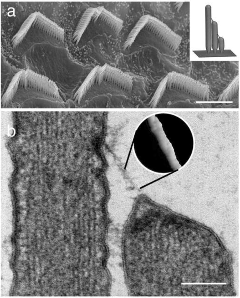
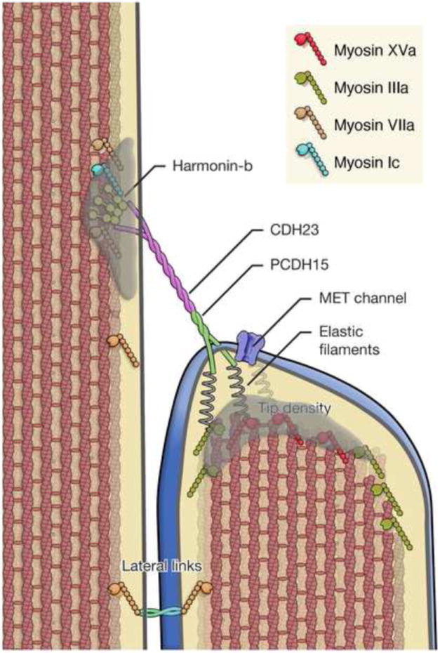
Recent findings: Structural, histological and biochemical studies show that the extracellular domains of two deafness-associated cadherins, cadherin 23 (CDH23) and protocadherin 15 (PCDH15), interact in trans to form the upper and lower part of each tip link, respectively. High-speed Ca imaging suggests that MET channels are localized exclusively at the lower end of each tip link. Biochemical and genetic studies provide evidence that defects in tip links cause hearing impairment in humans.

Summary: The identification of the proteins that form tip links have shed new light on the molecular basis of MET and the mechanisms causing hereditary deafness, noise-induced hearing loss and presbycusis.


Figures

Figure 1
Figure 1
Stereocilia of the inner ear hair cells form organized bundles and are connected to each other by tip links. (a) Scanning electron micrograph showing stereocilia bundles on the apical surface of the outer hair cells of the rat organ of Corti. Bar = 5 μ m. (Inset in a) Model depicting a longitudinal section through a hair cell bundle. Note the staircase pattern and arrangement of tip links. (b) Thin section transmission electron micrograph showing a tip link connecting one stereocilium to an adjacent taller neighbor. Note the presence of electron dense material at the upper and lower insertion sites and that the tip of the shorter stereocilia is tented due to tip link tension (from Kachar et al., 2000). Bar = 150 nm. (Inset in b) The surface rendering of a freeze-etching image of the tip link provides a close up view of helical structure of the tip link. (Adapted from [6] : Kachar B, Parakkal M, Kurc M et al.: High-resolution structure of hair-cell tip links. Proc Natl Acad Sci U S A 2000, 97:13336-41)
Figure 2
Figure 2
Schematic representation of the tip link complex illustrating structural features and key molecular components. CDH23 and PCDH15 comprise the tip link, which inserts into the stereocilia membrane at the sites of the upper and lower tip densities. Tip densities are presumed to contain scaffolding proteins, which bind to the cytoplasmic domain CDH23 and PCDH15 and anchor the tip link. In addition several myosins including myosin Ic, myosin IIIa, myosin VIIa, and myosin XVa have been localized to the tip density region and proposed to participate in MET adaptation, stereocilia actin dynamics, localization of lateral links, and cargo transport. Tension on the tip link gates the mechanoelectrical transduction (MET) channel. The tip-link can exert force onto the channel either directly or indirectly by tenting the membrane. The gating mechanism is presumed to involve a gating spring element through which force is applied to the channel gate. The channel gate opens in series with this spring, thus transiently reducing the force onto this spring and increasing compliance. The coiled structure of the tip link and the properties expected of conventional cadherins suggest that the tip link is relatively stiff. The spring element should then be in series with the tip link, possible as putative “elastic filaments” between the membrane and the dense actin core. Original figure

References

    1. Kazmierczak P, Sakaguchi H, Tokita J, et al. Cadherin 23 and protocadherin 15 interact to form tip-link filaments in sensory hair cells. Nature. 2007;449:87–91. - PubMed
    1. Siemens J, Lillo C, Dumont RA, et al. Cadherin 23 is a component of the tip link in hair-cell stereocilia. Nature. 2004;428:950–5. - PubMed
    1. Ahmed ZM, Goodyear R, Riazuddin S, et al. The tip-link antigen, a protein associated with the transduction complex of sensory hair cells, is protocadherin-15. J Neurosci. 2006;26:7022–34. - PMC - PubMed
    1. Ricci AJ, Kachar B, Gale J, et al. Mechano-electrical transduction: new insights into old ideas. J Membr Biol. 2006;209:71–88. - PMC - PubMed
    1. Pickles JO, Comis SD, Osborne MP. Cross-links between stereocilia in the guinea pig organ of Corti, and their possible relation to sensory transduction. Hear Res. 1984;15:103–12. - PubMed

Publication types

MeSH terms