

doi: 10.1038/nmeth.1349.
Epub 2009 Jul 26.
The twin spot generator for differential Drosophila lineage analysis
Affiliations
- PMID: 19633664
- PMCID: PMC2720837
- DOI: 10.1038/nmeth.1349
Item in Clipboard
The twin spot generator for differential Drosophila lineage analysis
Nat Methods.
2009 Aug.
Abstract
In Drosophila melanogaster, widely used mitotic recombination-based strategies generate mosaic flies with positive readout for only one daughter cell after division. To differentially label both daughter cells, we developed the twin spot generator (TSG) technique, which through mitotic recombination generates green and red twin spots that are detectable after the first cell division as single cells. We propose wide applications of TSG to lineage and genetic mosaic studies.
Figures
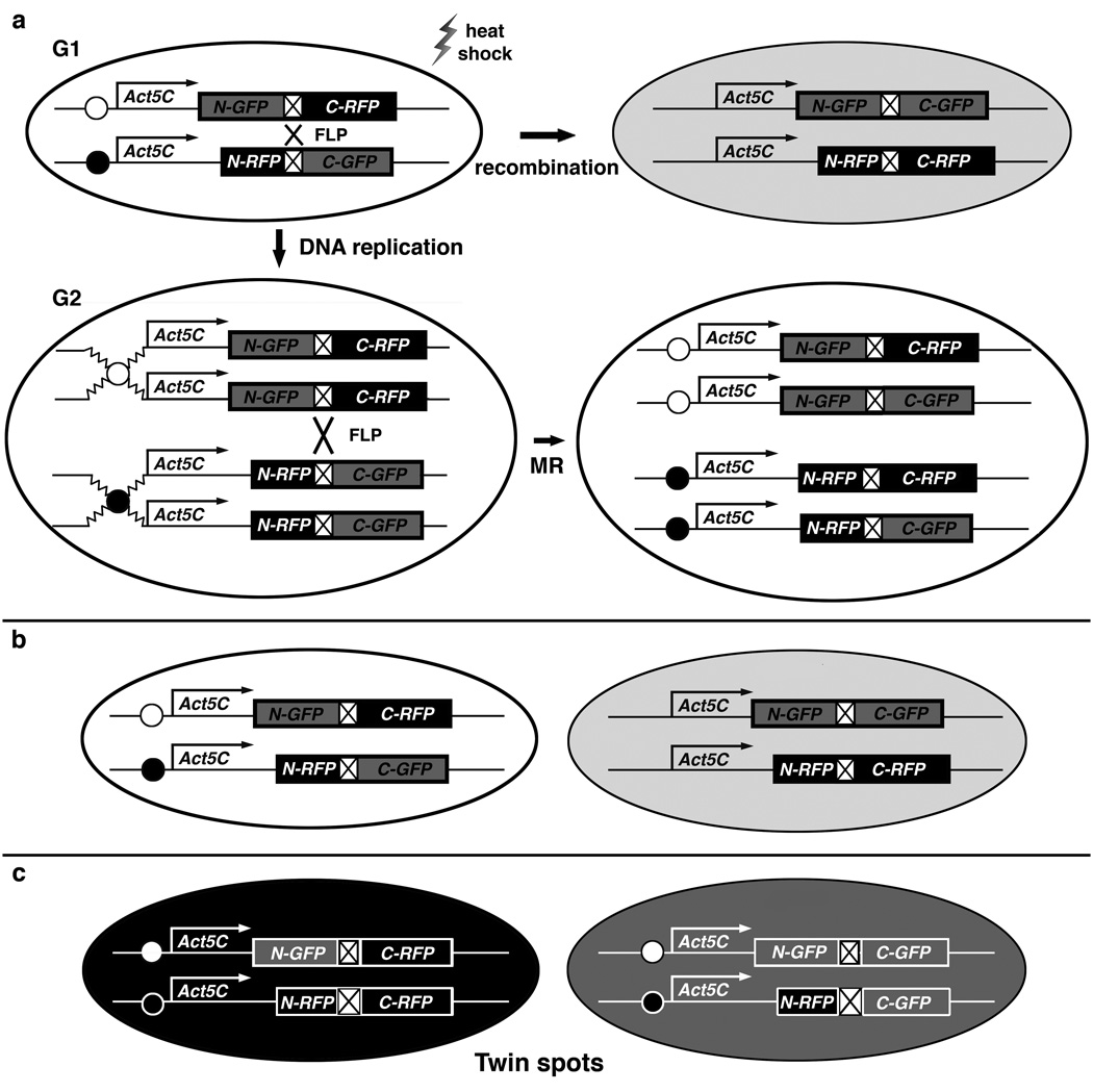
(a) Top: G1 recombination between homologous chromosomes generates two genotypically-identical yellow daughter cells expressing both GFP and RFP (Only one daughter cell is shown in light gray). Bottom left: duplicated chromosomes at G2. Bottom right: chromatids in cell just after mitotic recombination (MR). (b–c) MR occurs after DNA replication at G2. (b) In G2-Z segregation, recombinant chromosomes go to the same pole to generate a yellow daughter cell carrying both recombinant chromosomes, and a colorless daughter cell, carrying both non-recombinant chromosomes. (c) In G2-X segregation, recombinant chromosomes go to opposite poles to generate twin spots; that is, one red daughter cell, shown in black, and one green, shown in gray, each carrying one recombinant chromosome.
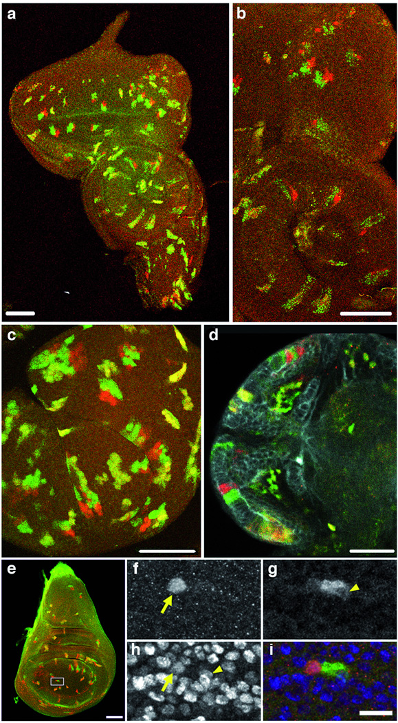
Red and green twin spots, and yellow clones, generated after MR at 82F7. (a–e) Bar scale: 50 um. (a–b) Initial GR-RG constructs, split at position 18 (Supplementary Methods). Eye-antennal imaginal disc. Dorsal up. Unstaged larvae: hs, 30–45 min, dissected at wandering third instar. (b) Enlargement reveals punctate GFP signal (Supplementary Table 1). (c–i) Final GR-RG constructs, split at position 349. GFP signal is homogeneous. (c) Haltere disc. Mid-third instar larvae: 30 min hs, dissected 24 h later. (d) Larval brain; anti-DsRed; anti-GFP; anti–DE-Cadherin stains the neuropil which gives rise to the optic lobe. Second instar larvae: 40 min hs, dissected 3–6 h later. (e) 2-cell clone in imaginal wing disc. Rectangle area enlarged in (f–i) Bar scale: 10 um. Yellow arrows point to one nucleus, and arrowheads to the other. (f) RFP expression. (g) GFP expression. (h) Nuclei stained with anti-histone. (i) Merged image.
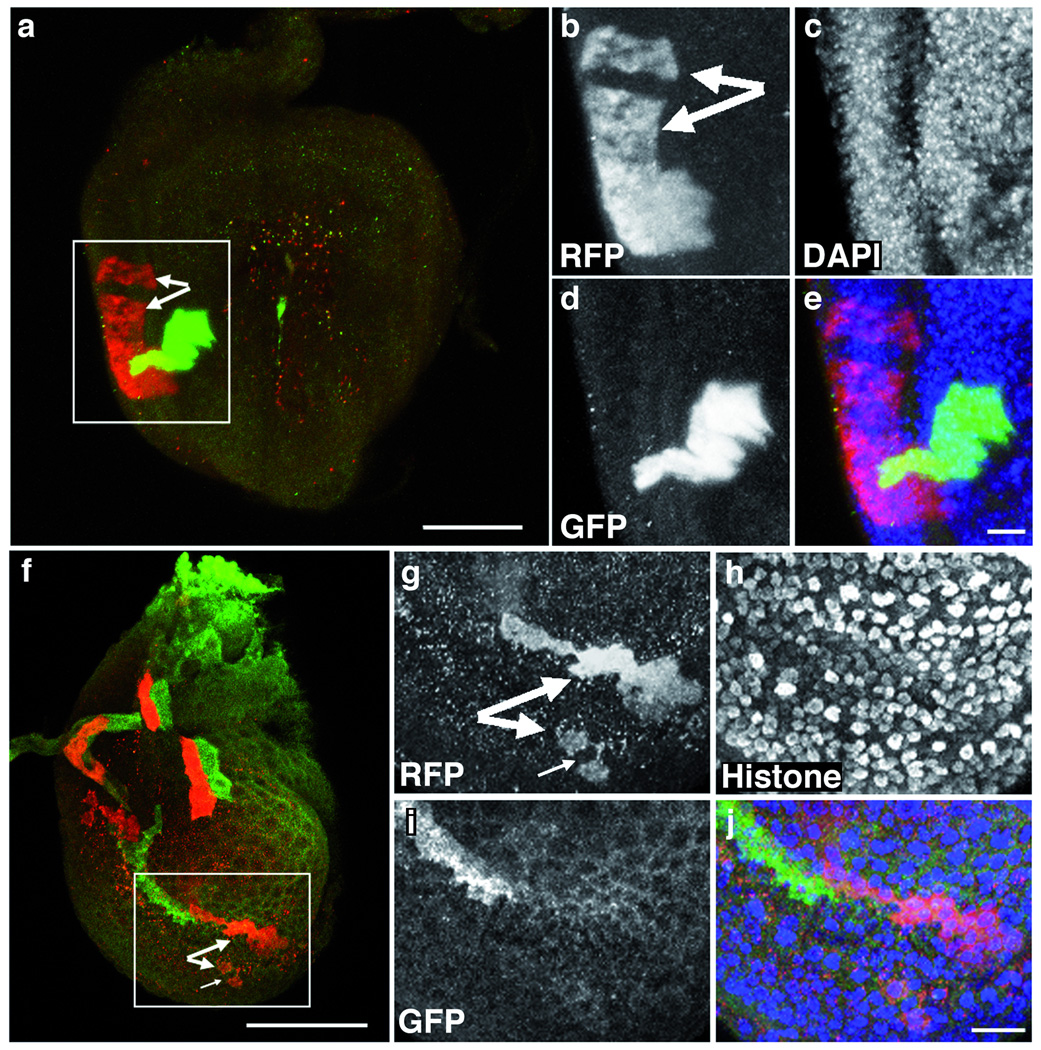
Projected z-series of late third-instar prothoracic leg discs with twin spots in: (a–e) the disc proper and (f–j) the peripodial epithelium. Dorsal is up. Bar scale is 50 µm in (a) and (f). We induced twin spots with MR at 82F7 with a 20 minute heat shock at 48 h AED, and fixed and stained discs at 120 h AED. We labeled samples in single-channel insets (b–d, g–i) with anti-DsRed to detect RFP (b, g), anti-GFP (d, i), and either DAPI (c) or anti-Histone (h) to mark nuclei. Big pairs of arrows (a, b, f, and g) indicate separated clones. The yellow color (a) is due to superposition of green/red clones in the projection. Small arrow (f, g) indicates an almost-separated clone. Merged images (e, j) demonstrate that clone separation is not due to damaged or missing cells. Bar scale is 10 µm in (e) and (j).
References
-
- Lee T, Luo L. Neuron. 1999;22:451–461. - PubMed
-
- Lee T, Luo L. Trends Neurosci. 2001;24:251–254. - PubMed
-
- de la Cova C, Abril M, Bellosta P, Gallant P, Johnston LA. Cell. 2004;117:107–116. - PubMed
-
- Spradling AC, Rubin GM. Science. 1982;218:341–347. - PubMed
-
- Zong H, Espinosa JS, Su HH, Muzumdar MD, Luo L. Cell. 2005;121:479–492. - PubMed
Publication types
MeSH terms
Grants and funding
LinkOut - more resources
Full Text Sources
Molecular Biology Databases
Research Materials
Miscellaneous
