. 2009 Aug;16(8):814-8.
doi: 10.1038/nsmb.1640. Epub 2009 Jul 26.

Role of mammalian Mre11 in classical and alternative nonhomologous end joining

Affiliations

Role of mammalian Mre11 in classical and alternative nonhomologous end joining

Anyong Xie et al. Nat Struct Mol Biol. 2009 Aug.

Abstract

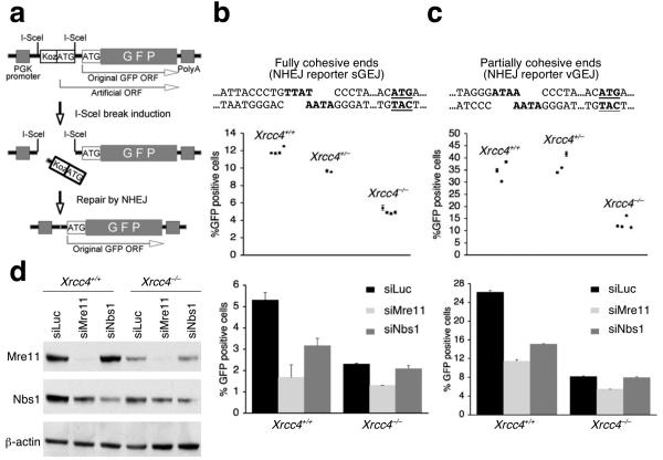
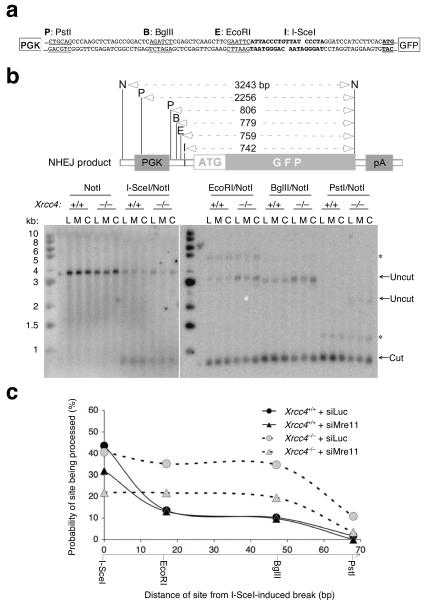
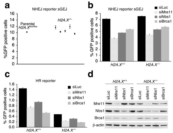
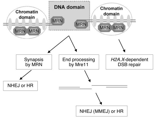
The mammalian Mre11-Rad50-Nbs1 (MRN) complex coordinates double-strand break signaling with repair by homologous recombination and is associated with the H2A.X chromatin response to double-strand breaks, but its role in nonhomologous end joining (NHEJ) is less clear. Here we show that Mre11 promotes efficient NHEJ in both wild-type and Xrcc4(-/-) mouse embryonic stem cells. Depletion of Mre11 reduces the use of microhomology during NHEJ in Xrcc4(+/+) cells and suppresses end resection in Xrcc4(-/-) cells, revealing specific roles for Mre11 in both classical and alternative NHEJ. The NHEJ function of Mre11 is independent of H2A.X. We propose a model in which both enzymatic and scaffolding functions of Mre11 cooperate to support mammalian NHEJ.


Figures

Figure 1
Figure 1
Mre11 regulates both Xrcc4-dependent and Xrcc4-independent NHEJ. (a) Structure of the NHEJ reporter. PGK: phosphoglycerate kinase. “Koz-ATG”: an artificial Kozak-ATG translation start site. ORF: open reading frame. PolyA: polyadenylation signal. (b, c) I-SceI-induced NHEJ in Xrcc4+/+ and Xrcc4−/− isogenic mouse ES cells containing the sGEJ reporter (b) or the vGEJ reporter (c). In upper sections, each point represents the mean of triplicates in one experiment for each reporter clone; error bars indicate standard error of mean (s.e.m). Unpaired t-test (unknown variance) for Xrcc4−/− cells versus Xrcc4+/+ cells, P = 0.00000037 (b) or P = 0.0038 (c); for Xrcc4−/− cells versus Xrcc4+/− cells, P = 0.000015 (b) or P = 0.0025 (c). In lower sections, Xrcc4+/+ and Xrcc4−/− NHEJ reporter cells were co-transfected with siRNA to Mre11 (siMre11), Nbs1 (siNbs1), or control luciferase (siLuc), together with I-SceI expression plasmids. Percentages of I-SceI-induced GFP+ cells were measured. Bars represent the mean of triplicates in one representative experiment. Error bars indicate s.e.m. Student's paired t-test (two-tailed): in Xrcc4+/+ cells, siMre11 versus siLuc, P = 0.00045 (b) and P = 0.000035 (c); siNbs1 versus siLuc, P = 0.0037 (b) and P = 0.0012 (c); in Xrcc4−/− cells, siMre11 versus siLuc, P = 0.0037 (b) and P = 0.0018 (c); siNbs1 versus siLuc, P = 0.07 (b) and P = 0.467 (c). (d) Protein abundance in Xrcc4+/+ and Xrcc4−/− NHEJ reporter mouse ES cells treated with siRNAs shown. Whole cell extracts were analyzed three days after siRNA transfection. β-actin is a loading control.
Figure 2
Figure 2
Mre11 promotes end processing in Xrcc4-independent NHEJ.(a) Junction sequence of the NHEJ reporter sGEJ “pop-out” product (i.e., with excision of the Kozak-ATG translation start site). Sequences of two partial I-SceI sites are indicated in bold. Start codon of the GFP ORF is in bold and underlined. Restriction sites in the reporter are as follows. P: PstI; B: BglII; E: EcoRI; I: I-SceI. PGK: phopshoglycerate kinase promoter. (b) Structural analysis of pooled I-SceI-induced GFP+ NHEJ products. Upper section shows sizes of expected GFP-hybridizing restriction fragment sizes following digestion of gDNA with enzymes shown (I: I-SceI; E: EcoRI; B: BglII; P; PstI; N: NotI). Lower section shows Southern blot analysis of gDNA from pooled I-SceI-induced GFP+ NHEJ products from Xrcc4+/+ and Xrcc4−/− sGEJ reporter mouse ES cells co-transfected with I-SceI expression vector and either control siLuc (L) or test siMre11 (M) or siCtIP (siRNA to CtIP) (C). Southern blots were probed with GFP cDNA. Arrows indicate NHEJ products that had either retained (“Cut”) or lost (“Uncut”) the relevant I-SceI-proximal restriction site. PGK: phopshoglycerate kinase promoter. pA: polyadenylation signal. *: non-specific hybridization product. (c) Effect of Xrcc4 status and Mre11 depletion on the probability of restriction sites being processed during NHEJ, calculated from the intensities of the DNA bands in the Southern blot in (b), quantified by phosphoimager and densitometry, as the intensity of the “uncut” band divided by the combined intensities of “cut” and “uncut” bands (expressed as a percentage). The probability is plotted against the distance of each site from the I-SceI-induced break indicated along the x-axis.
Figure 3
Figure 3
The HR and NHEJ function of Mre11 is at least in part independent of H2A.X. (a) I-SceI-induced NHEJ in parental H2A.X+/+ and isogenic H2A.X−/− sGEJ NHEJ reporter mouse ES clones. Points represent mean of triplicates for independent clones. Error bars indicate s.e.m. (b, c) Percentage of I-SceI-induced GFP+ cells from H2A.X+/+ and H2A.X−/− sGEJ reporter mouse ES cells (b) or HR reporter mouse ES cells (c) depleted of Mre11, Nbs1 or Brca1 by siRNA duplex, with siLuc as a control. Bars represent mean of triplicates. Error bars indicate s.e.m. Student's paired t-test (two-tailed) in (b): in H2A.X+/+ cells, siLuc versus siMre11, P = 0.000048; versus siNbs1, P = 0.0009; versus siBrca1, P = 0.0024; in H2A.X−/− cells, siLuc versus siMre11, P = 0.00089; versus siNbs1, P = 0.0013; versus siBrca1, P = 0.0046. Student's paired t-test in (c): in H2A.X+/+ cells, siLuc versus siMre11, P = 0.0023; versus siNbs1, P = 0.0046; versus siBrca1, P = 0.00026; in H2A.X−/− cells, siLuc versus siMre11, P = 0.00055; versus siNbs1, P = 0.0059; versus siBrca1, P = 0.000043. (d) Steady state protein levels in H2A.X+/+ and H2A.X−/− reporter mouse ES cells treated with indicated siRNAs. Whole cell extracts were analyzed by Western blotting three days after siRNA transfection. β-actin serves as a loading control.
Figure 4
Figure 4
Model for MRN's functions at a mammalian DSB. Mre11 nuclease activity may resect the DNA ends for either HR or Xrcc4-independent NHEJ/MMEJ. MRN may also promote synapsis during NHEJ (and possibly during HR). “Short-range” NHEJ of repairable DSBs by MRN appears to be independent of H2A.X.

Comment in

References

    1. Williams RS, Williams JS, Tainer JA. Mre11-Rad50-Nbs1 is a keystone complex connecting DNA repair machinery, double-strand break signaling, and the chromatin template. Biochem Cell Biol. 2007;85:509–20. - PubMed
    1. Berkovich E, Monnat RJ, Jr., Kastan MB. Roles of ATM and NBS1 in chromatin structure modulation and DNA double-strand break repair. Nat Cell Biol. 2007;9:683–90. - PubMed
    1. Lee JH, Paull TT. ATM activation by DNA double-strand breaks through the Mre11-Rad50-Nbs1 complex. Science. 2005;308:551–4. - PubMed
    1. Stucki M, Jackson SP. gammaH2AX and MDC1: anchoring the DNA-damage-response machinery to broken chromosomes. DNA Repair (Amst) 2006;5:534–43. - PubMed
    1. Bekker-Jensen S, et al. Spatial organization of the mammalian genome surveillance machinery in response to DNA strand breaks. J Cell Biol. 2006;173:195–206. - PMC - PubMed

Publication types

MeSH terms