. 2009 Aug;16(8):808-13.
doi: 10.1038/nsmb.1639. Epub 2009 Jul 26.

Multiple functions of MRN in end-joining pathways during isotype class switching

Affiliations

Multiple functions of MRN in end-joining pathways during isotype class switching

Maria Dinkelmann et al. Nat Struct Mol Biol. 2009 Aug.

Abstract

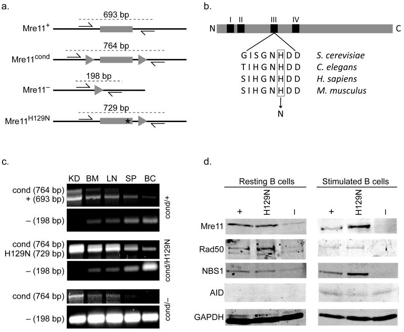
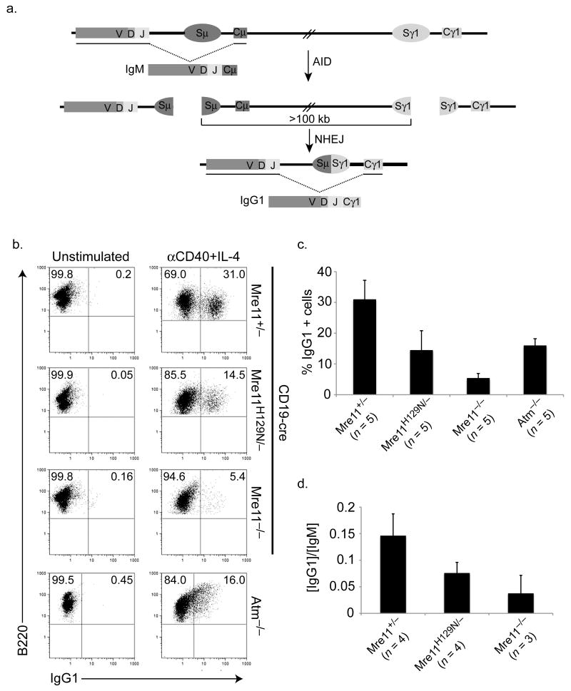
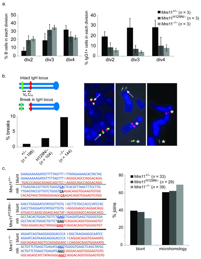
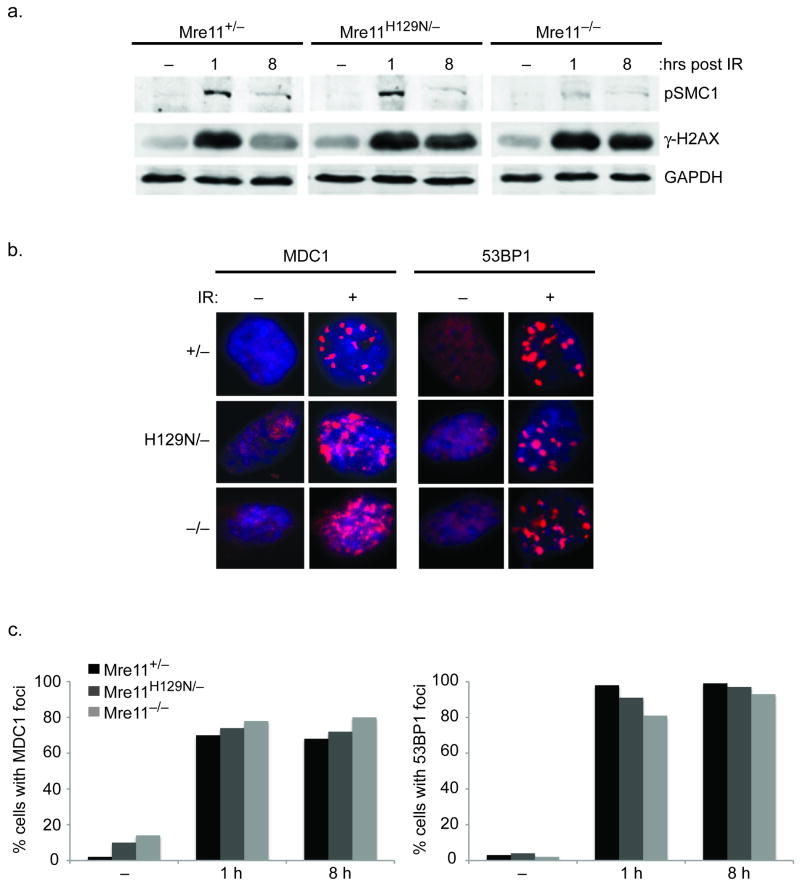
The Mre11-Rad50-NBS1 (MRN) complex has many roles in response to DNA double-strand breaks, but its functions in repair by nonhomologous end joining (NHEJ) pathways are poorly understood. We have investigated requirements for MRN in class switch recombination (CSR), a programmed DNA rearrangement in B lymphocytes that requires NHEJ. To this end, we have engineered mice that lack the entire MRN complex in B lymphocytes or that possess an intact complex that harbors mutant Mre11 lacking DNA nuclease activities. MRN deficiency confers a strong defect in CSR, affecting both the classic and the alternative NHEJ pathways. In contrast, absence of Mre11 nuclease activities causes a milder phenotype, revealing a separation of function within the complex. We propose a model in which MRN stabilizes distant breaks and processes DNA termini to facilitate repair by both the classical and alternative NHEJ pathways.


Conflict of interest statement

The authors declare no competing financial interests.

Figures

Figure 1
Figure 1. Mre11 deficiencies in the B lymphocyte lineage
(a) Four germline mouse alleles of Mre11: Mre11 wild type (Mre11+), Mre11 conditional (Mre11cond), Mre11 null (Mre11), and Mre11 deficient in nuclease activities (Mre11H129N). Mre11 exon 5 (gray rectangle), intronic sequence (black line), LoxP sites (triangles), histidine to asparagine mutation at amino acid 129 (asterisk). Base pair (bp) numbers indicate allele specific PCR products resulting from the two primers depicted (arrows). (b) Location of the invariant active site histidine within nuclease motif III. Mouse histidine 129 was changed to asparagine as described. (c) PCR analyses distinguishing the four Mre11 germline alleles. Conversion of Mre11cond to Mre11 is detected only in sites containing substantial numbers of B lymphocytes (BM - bone marrow, LN - lymph node, SP - whole spleen, BC - enriched B cell population from spleen), but not in kidney (KD). Primers used depicted in (a). (d) Western blot analyses of MRN components and AID in splenic B lymphocytes from mice harboring one allele of CD19-Cre. GAPDH was used as a protein loading control. Left - resting B cells, right - B cells stimulated to undergo class switch recombination for 4 days in culture. Each mouse contains the Mre11 allele indicated, and a second allele which is Mre11 in the B lineage (Mre11cond elsewhere).
Figure 2
Figure 2. Distinct deficiencies in isotype switching conferred by MRN and ATM mutations
(a) Schematic of class switching from default IgM to the IgG1 isotype. AID initiates the formation of DSBs in switch regions (Sμ and Sγ1) upstream of their corresponding constant regions (Cμ and Cγ1). The intervening sequence (>100 kb) is excised, and the remaining DNA ends are ligated by NHEJ pathways generating a new heavy chain gene encoding Cγ1. Schematic is not to scale. (b) Flow cytometric analyses of class switching from IgM to IgG1 in B cells cultured with IL-4 and anti-CD40 for four days. Numbers in upper right quadrants represent the average percentage of IgG1+ cells from three to five mice of each genotype. (c) Bar graph depicting direct comparisons of IgG1+ cell populations in (b) (average +/- S.D). (d) Antibodies secreted by stimulated B cells after 4 days in culture. Y axis indicates the ratio of IgG1 to IgM. No difference in IgM levels were detected (not shown).
Figure 3
Figure 3. Requirement for MRN in the repair process
(a) Reduced class switching in MRN deficiencies does not result from proliferation defects. Left - The cell tracking dye CFSE was used to distinguish populations of stimulated B cells having undergone the indicated number of divisions (x axis). The percentage of cells in each population is shown on the Y axis. Reduced proliferation in the Mre11 deficiencies is evident. Right - the percentage of cells in each population (x axis) having switched to IgG1. Reduced switching is evident in each population. (b) Chromosome breakage in the IgH locus. Two color FISH with BACs flanking IgH on mouse chromosome 12 reveals IgH breaks by separation of red (BAC 199) and green (BAC 207) signals. The representative examples show a normal metaphase (left) and two with an IgH break (center and right). Arrows indicate co-localized signals (intact). Asterisks indicate separated signals (broken). The bar graph depicts the percentage of metaphases with an IgH break. Three mice of each genotype were analyzed. The total number of metaphases analyzed are indicated below each genotype. (c) Mre11 deficiencies impact the Classic and Alternative end joining pathways. Left - Representative sequences of cloned joins involving the mu and gamma 1 switch regions. Right - The distribution of joins containing blunt ends (Lig4/XRCC4 dependant, classic NHEJ) and microhomologies (Lig4/XRCC4 independent, alternative NHEJ) are shown. Three mice of each genotype were analyzed. The total number of sequenced joins are indicated.
Figure 4
Figure 4. Independence of γ-H2AX-MDC1-53BP1 and MRN
(a) Distinct requirements for phosphorylation of SMC1 versus H2AX during the response to DSBs. Western blot analyses of the indicated proteins (right) prior to 10 Gy IR (−), and 1 or 8 hours post-IR. Phosphorylated SMC1 is used as a control to demonstrate functional MRN deficiency. GAPDH is a protein loading control. (b) Representative immunoflourescent foci prior to (−), or 8 hours after (+) 10 Gy IR. (c) The formation of MDC1 and 53BP1 foci at 1 and 8 h post-IR does not require Mre11 nuclease activities or MRN. Bar graphs represent quantification of MDC1 and 53BP1 foci-positive cells at the indicated times (X axis) after 10 Gy IR. (−) no IR. Positive cells defined as > 5 foci.

Comment in

References

    1. McKinnon PJ, Caldecott KW. DNA strand break repair and human genetic disease. Annu Rev Genomics Hum Genet. 2007;8:37–55. - PubMed
    1. Wyman C, Kanaar R. DNA double-strand break repair: all's well that ends well. Annu Rev Genet. 2006;40:363–83. - PubMed
    1. Audebert M, Salles B, Calsou P. Involvement of poly(ADP-ribose) polymerase-1 and XRCC1/DNA ligase III in an alternative route for DNA double-strand breaks rejoining. J Biol Chem. 2004;279:55117–26. - PubMed
    1. Corneo B, et al. Rag mutations reveal robust alternative end joining. Nature. 2007;449:483–6. - PubMed
    1. Soulas-Sprauel P, et al. Role for DNA repair factor XRCC4 in immunoglobulin class switch recombination. J Exp Med. 2007;204:1717–27. - PMC - PubMed

Publication types

MeSH terms

Substances