. 2009 Sep;10(9):1000-7.
doi: 10.1038/ni.1774. Epub 2009 Jul 26.

Instability of the transcription factor Foxp3 leads to the generation of pathogenic memory T cells in vivo

Affiliations

Instability of the transcription factor Foxp3 leads to the generation of pathogenic memory T cells in vivo

Xuyu Zhou et al. Nat Immunol. 2009 Sep.

Abstract

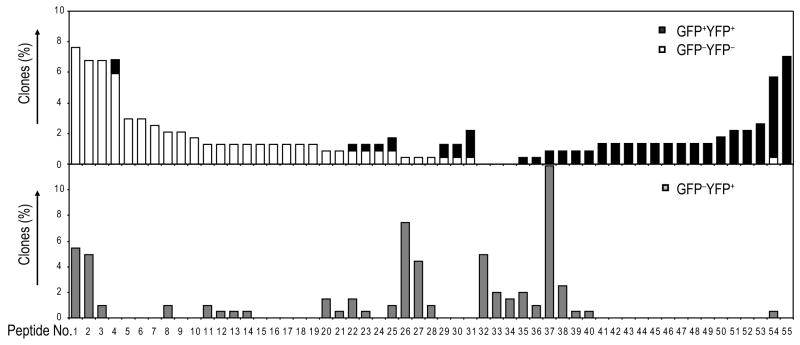
Regulatory T cells (T(reg) cells) are central to the maintenance of immune homeostasis. However, little is known about the stability of T(reg) cells in vivo. In this study, we demonstrate that a substantial percentage of cells had transient or unstable expression of the transcription factor Foxp3. These 'exFoxp3' T cells had an activated-memory T cell phenotype and produced inflammatory cytokines. Moreover, exFoxp3 cell numbers were higher in inflamed tissues in autoimmune conditions. Adoptive transfer of autoreactive exFoxp3 cells led to the rapid onset of diabetes. Finally, analysis of the T cell receptor repertoire suggested that exFoxp3 cells developed from both natural and adaptive T(reg) cells. Thus, the generation of potentially autoreactive effector T cells as a consequence of Foxp3 instability has important implications for understanding autoimmune disease pathogenesis.


Figures

Fig. 1
Fig. 1. Development of Foxp3+ T cells inFoxp3-GFP-Cre × R26-YFPtransgenic mice
(A)Thymocytes were isolated, stained for CD4 and CD8, and GFP and YFP expression was analyzed by flow cytometry. Thymocytes were gated as CD4+CD8+ double positive (DP) and individual CD4 or CD8 single positive (SP) populations. (B) The histograms show CD24 expression on the CD4SP GFP and YFP populations gated as shown in panel A. (C) GFP and YFP expression by gated CD4+ T cells from lymph nodes (LN) and spleen. (D) Methylation analysis of the Foxp3 locus. The methylation status of the CpG motifs of the TSDR of sub-cloned from purified Tconv, Tregs and exFoxp3 cells. Data is the average of data generated in3 independent experiments. (E) Graphs show the indicated GFP and YFP populations as a percentage of CD4+ T cells in spleen, LN, liver and Peyer’s patch of Foxp3-GFP-CrexR26-YFP mice. Each point represents an individual mouse. Experiments were performed with 6 -9 week old mice and are representative of 4 (A, B) and 20 (C) independent experiments.
Fig. 2
Fig. 2. CD4+ YFP+ Foxp3 cells have a non-Treg surface phenotype
(A) Lymph node and spleen CD4+ GFP+ T cells were FACS purified from Foxp3-GFP-Cre mice and CD4+ YFP+ T cells sort-purified from Foxp3-GFP-Crex R26-YFPmice. Purified populations were analyzed for CD127, CD25, and GITR and Foxp3 by intracellular staining. (B) As in (A), analysis of FR4, CTLA-4 and CD103 expression and Foxp3 by intracellular staining. Histograms show YFP Tconv (thick line), YFP+ Foxp3+ Treg (thin line), and YFP+ Foxp3 (filled histogram). Representative plots from 6–9 week old mice of 3 experiments.
Fig. 3
Fig. 3. CD4+ YFP+ Foxp3 cells have a non-Treg, memory cell surface phenotype and produce IFN-γ and IL-17
Analysis of peripheral lymphoid organ resident CD4+ GFP+YFP+, CD4+ GFP YFP+, and CD4+GFPYFP cells. (A) Spleen CD4+ populations were gated and analyzed for CD62L and CD44 expression. (B) Spleen CD4+ YFP+ and CD4+ YFP T cells were purified then stimulated with PMA, ionomycin and monensin for 4 h; cells were then stained for intracellular Foxp3 and IFN-γ. (C,D) Graphs show the percent of cells in each population that produce IFN-γ (C) or IL-17 (D). Each point represents an individual mouse. Experiments were performed on 6 – 9 week old mice and are representative of 5 (A, B) independent experiments.
Fig. 4
Fig. 4. The autoimmune microenvironment favors loss of Foxp3
(A)Percent of GFPYFP+ exFoxp3 cells among CD4+YFP+ cells in the pancreas, inguinal (ILN) and pancreatic (PLN) lymph nodes of NOD mice. Data points represent individual mice. (B,C) CD25 (B) orCD127 (C) expression on exFoxp3 cells isolated from each site, expressed as mean fluorescence intensity (MFI). Six 16–18-week old mice were analyzed in 2 independent experiments. (D) Development of Treg and exFoxp3 cells in BDC2.5 TCR Tg mice. Analysis of GFP and YFP expression in thymocytes, LN cells and splenocytes of 6–9 week-old BDC2.5 TCR Tg+ and control TCR Tg mice. Plots are gated on live thymocytes and enriched CD4+ T cells from LN and spleen. Representative of 3 mice. (E) CD4+ YFP+ T cells purified from pancreatic LN (PLN) of BDC2.5.TCR Tg or non-Tg control mice were analyzed for Vβ4 (BDC2.5 TCR chain) and Foxp3 expression. Plots are representative of 3 mice, and each point on the graph represents the percentage of YFP+ FoxP3 cells in individual mice.
Fig. 5
Fig. 5. exFoxp3 cells develop from Treg upon adoptive transfer
(A)GFP+YFP+ GITRhigh BDC2.5 TCR Tg purified cells (left dot plot) were transferred into NOD Tcra−/− mice. After four weeks splenic YFP+ cells were purified, exposed to PMA, ionomycin and monensin for 3–4 hours and stained for intracellular Foxp3 and IFN-γ (right dot plot). Representative of 4 mice. (B) GFPYFP cells purified from inguinal LNs (ILN) of BDC2.5 TCR Tg+ mice were transferred into NOD Rag2−/− mice. Seven days after transfer, pancreatic mononuclear cells of acutely diabetic NOD Rag2−/− mice that received CD4+ BDC2.5 TCR Tg+ YFP cells, or of control NOD Rag2−/− mice that did not receive cells were analyzed by flow cytometry. CD4+ Vβ4+ cells were gated and analyzed for YFP expression. Representative of 3 mice.
Fig. 6
Fig. 6. exFoxp3 cells are pathogenic
(A)CD4+ T cells from Foxp3-GFP-Cre × R26-YFPBDC2.5 mice were sorted to >95% purity into GFP+YFP+ and GFPYFP+ cells. The percentage of Foxp3+ within these sorted populations was determined using intracellular staining (pre-culture). (B) Purified cells were cultured withanti-CD3 and anti-CD28 coated beads + IL-2for 6–8days, and were stained by flow cytometry (post-culture). Representative plots from 3 experiments.(C) 5 × 10⁵ expanded GFPYFP, GFP+YFP+ or GFPYFP+ cells were transferred into NOD Rag2−/− mice, and blood glucose concentrations were recorded from 8 days later and onwards. Diabetes is considered when blood glucose exceeds 250 mg/dl. Each point represents an individual mouse. (D) Hematoxylin and eosin staining of pancreas sections from NOD Rag2−/− mice that did not receive cells, or that received GFPYFP+ cells 9 days prior as described in (B). Representative of 4 mice.
Fig. 7
Fig. 7. exFoxp3 cells can develop from both nTregs and aTregs
CD4+ GFPYFP, GFPYFP+ and GFP+YFP+ T cells were sorted from BDC2.5 TCR Tg mice, cDNA was amplified with Vα2-specific primers and the amplicons were subcloned and sequenced. The frequency of each unique CDR3 amino acid sequence in Tconv (un-filled bar), exFoxp3 (grey bar) and Treg cells (black bar). Data is one mouse representative of 2 analysed.

Comment in

References

    1. Tang Q, et al. Central role of defective interleukin-2 production in the triggering of islet autoimmune destruction. Immunity. 2008;28:687–97. - PMC - PubMed
    1. Bluestone JA, Tang Q, Sedwick CE. T regulatory cells in autoimmune diabetes: past challenges, future prospects. J Clin Immunol. 2008;28:677–84. - PubMed
    1. Wan YY, Flavell RA. Regulatory T-cell functions are subverted and converted owing to attenuated Foxp3 expression. Nature. 2007;445:766–70. - PubMed
    1. Hori S, Nomura T, Sakaguchi S. Control of regulatory T cell development by the transcription factor Foxp3. Science. 2003;299:1057–61. - PubMed
    1. Bennett CL, Ochs HD. IPEX is a unique X-linked syndrome characterized by immune dysfunction, polyendocrinopathy, enteropathy, and a variety of autoimmune phenomena. Curr Opin Pediatr. 2001;13:533–8. - PubMed

Publication types

MeSH terms