. 2009 Jul 27;4(7):e6321.
doi: 10.1371/journal.pone.0006321.

Characterization of the PMT gene family in Cryptococcus neoformans

Affiliations

Characterization of the PMT gene family in Cryptococcus neoformans

Sven D Willger et al. PLoS One. .

Abstract

Background: Protein-O-mannosyltransferases (Pmt's) catalyze the initial step of protein-O-glycosylation, the addition of mannose residues to serine or threonine residues of target proteins.

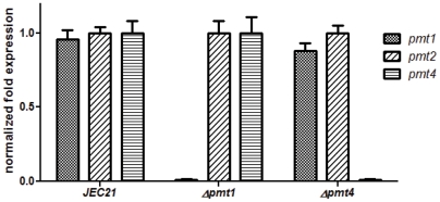
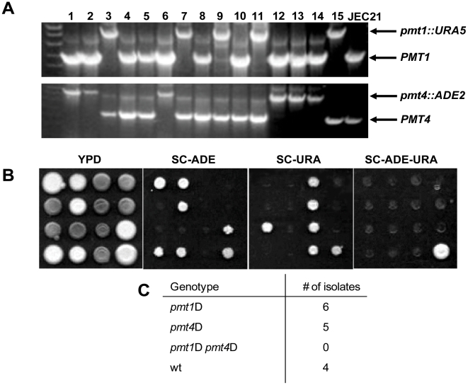
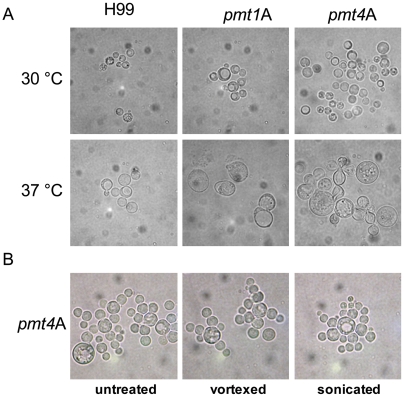
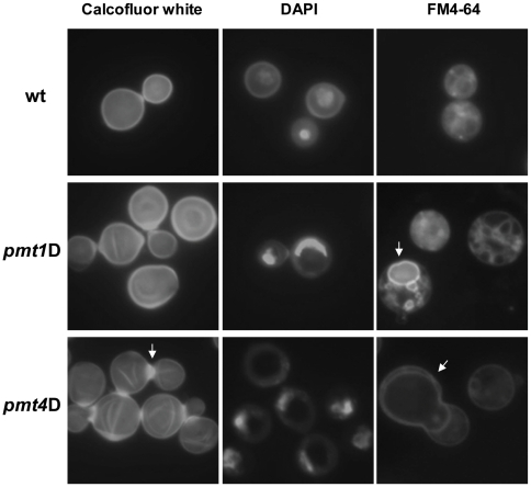
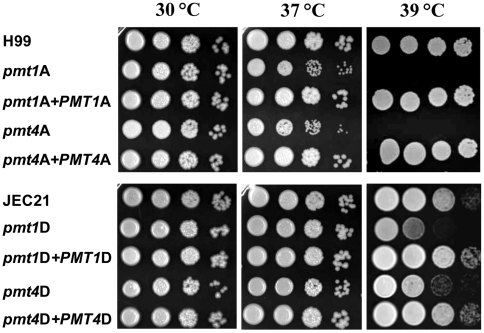
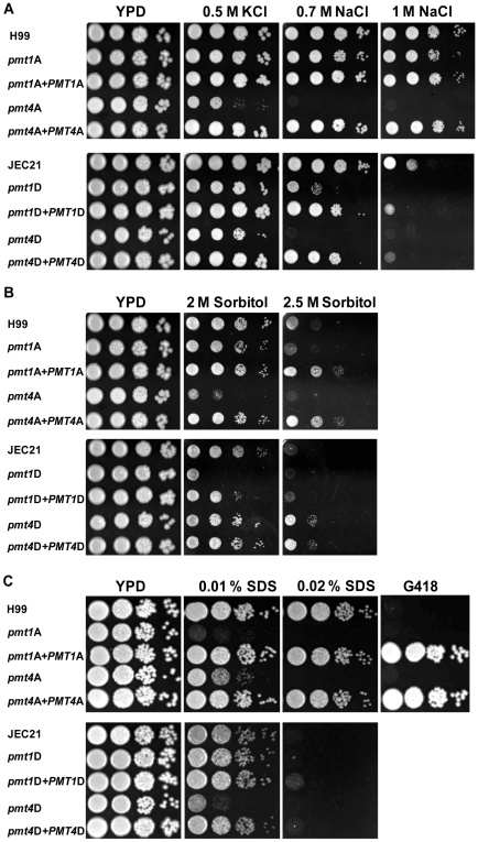
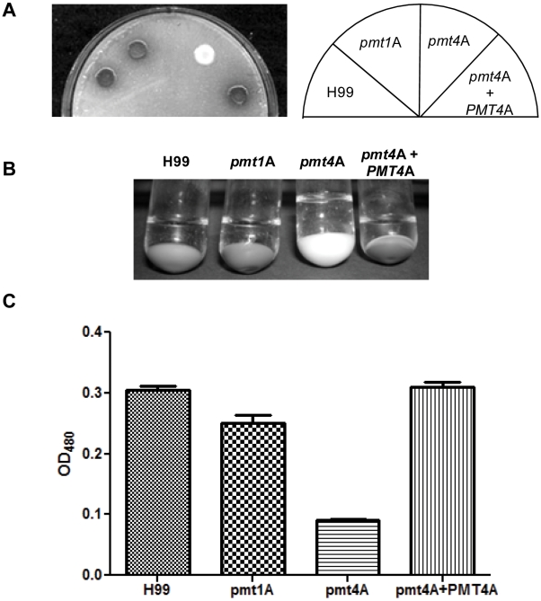
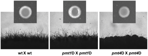
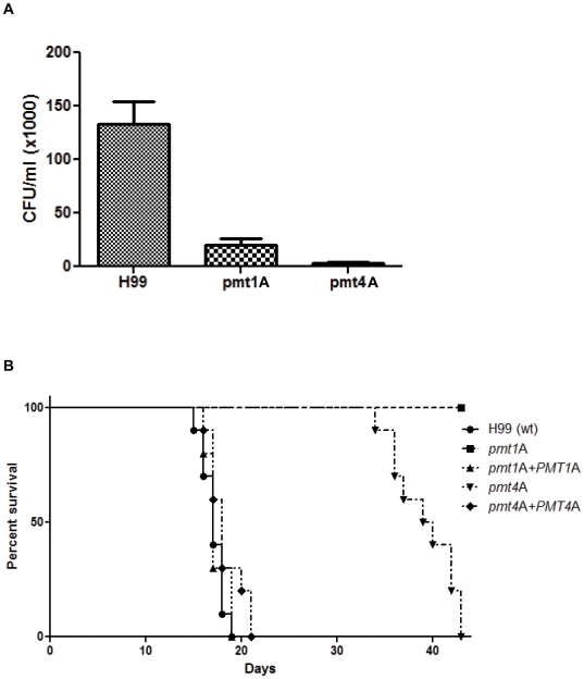
Methodology/principal findings: Based on protein similarities, this highly conserved protein family can be divided into three subfamilies: the Pmt1 sub-family, the Pmt2 sub-family and the Pmt4 sub-family. In contrast to Saccharomyces cerevisiae and Candida albicans, but similar to filamentous fungi, three putative PMT genes (PMT1, PMT2, and PMT4) were identified in the genome of the human fungal pathogen Cryptococcus neoformans. Similar to Schizosaccharomyces pombe and C. albicans, C. neoformans PMT2 is an essential gene. In contrast, the pmt1 and pmt4 single mutants are viable; however, the pmt1/pmt4 deletions are synthetically lethal. Mutation of PMT1 and PMT4 resulted in distinct defects in cell morphology and cell integrity. The pmt1 mutant was more susceptible to SDS medium than wild-type strains and the mutant cells were enlarged. The pmt4 mutant grew poorly on high salt medium and demonstrated abnormal septum formation and defects in cell separation. Interestingly, the pmt1 and pmt4 mutants demonstrated variety-specific differences in the levels of susceptibility to osmotic and cell wall stress. Delayed melanin production in the pmt4 mutant was the only alteration of classical virulence-associated phenotypes. However, the pmt1 and pmt4 mutants showed attenuated virulence in a murine inhalation model of cryptococcosis.

Conclusion/significance: These findings suggest that C. neoformans protein-O-mannosyltransferases play a crucial role in maintaining cell morphology, and that reduced protein-O-glycosylation leads to alterations in stress resistance, cell wall composition, cell integrity, and survival within the host.


Conflict of interest statement

Competing Interests: The authors have declared that no competing interests exist.

Figures

Figure 1
Figure 1. PMTs are not differentially expressed and the loss of one pmt gene is not compensated by overexpression of another PMT gene.
The wild-type (JEC21), Δpmt1 and Δpmt4 mutant strains were incubated to mid-logarithmic phase in YPD medium and total RNA was isolated. Quantitative real-time PCR was performed on the corresponding cDNA samples to assess relative PMT1, PMT2, and PMT4 transcript abundance compared to the wild-type strain±one standard deviation. The expression values in all three strains are normalized to the PMT2 expression.
Figure 2
Figure 2. A pmt1 pmt4 double deletion is lethal in serotype D.
Serotype D strains pmt1D (MATa pmt1D::URA5 ade2-27) and pmt4D (MATα pmt4D::ADE2 ura5) were crossed on standard V8 mating media and individual spores were isolated after several days by micromanipulation. A: 15 individual progeny were analyzed by colony PCR for the presence of the PMT1 (upper panel) and PMT4 alleles (lower panel). Position of the respective wild-type and disruption alleles are indicated at right. Wild-type strain JEC21 was used as a control. B: The 15 strains from A were spotted onto the indicated plates, grown for 2–3 days at 30°C and subsequently analyzed for auxotrophic marker distribution. Strains were spotted from top left (#1) to bottom right (wild-type control). C: Summary of the genotypes identified in A and B with respect to PMT alleles and mating-type. Mating-type of the individual spores were determined by standard mating reactions using wild-type strains JEC20 (MATa) and JEC21 (MATα) as tester strains.
Figure 3
Figure 3. The pmt1 and pmt4 disruption strains show altered cell morphology.
A: Serotype A wild-type strain H99 and mutant strains pmt1A (pmt1A::URA5) and pmt4A (pmt4A::URA5) were incubated in YPD at 30°C and 37°C to an OD600 of 1, and cells were subsequently analyzed by light microscopy (DIC). B: Serotype A pmt4A strain was incubated in YPD at 30°C to an OD600 of 1, and cell suspension was analyzed by light microscopy (DIC) after no treatment or 1 min vortexing or sonication.
Figure 4
Figure 4. pmt1 and pmt4 mutant strains show altered cell wall phenotypes and vacuole distribution.
Wild-type serotype A strain H99 and pmt mutant strains pmt1A (pmt1A::URA5) and pmt4A (pmt4A::URA5) were incubated in YPD at 30°C to an OD600 of 1. Cells were fixed for 30 min in 10% formaldehyde and subsequently stained as described with chitin staining dye calcofluor white, the DNA dye DAPI and the vacuolar dye FM4-64. Pictures were taken at 400x using a Zeiss Axioskop 2 Plus Fluorescence Microscope and a AxioCam MRM digital camera.
Figure 5
Figure 5. pmt mutant strains show growth defects at high growth temperatures.
Over-night cultures of wild-type, pmt mutant and corresponding reconstituted strains from serotype A (upper panel) and serotype D (lower panel) were adjusted to an OD600 of 0.1 and diluted by 10-fold serial dilutions down to a 10−4 dilution. 5 µl of each dilution step was spotted onto YPD plates, and plates were incubated at the indicated temperature for 2–3 days. Strains used were the serotype A strains H99 (wild-type), pmt1A (pmt1A::URA5), pmt4A (pmt4A::URA5), pmt1A+PMT1A (pmt1A::URA5 PMT1A-NeoR) and pmt4A+PMT4A (pmt4A::URA5 PMT4A-NeoR), and the respective serotype D strains JEC21 (wild-type), pmt1D (pmt1D::URA5), pmt4D (pmt4D::URA5), pmt1D+PMT1D (pmt1D::URA5 PMT1D-NeoR) and pmt4D+PMT4D (pmt4D::URA5 PMT4D-NeoR).
Figure 6
Figure 6. pmt mutant strains are sensitive to hyper-osmotic stresses.
Over-night cultures of wild-type, pmt mutant and corresponding reconstituted strains from serotype A (upper panel) and serotype D (lower panel) were adjusted to an OD600 of 0.1 and diluted by 10-fold serial dilutions down to a 10−4 dilution. 5 µl of each dilution step was spotted onto YPD plates containing stress agents, and plates were incubated at the indicated temperature for 2–3 days. Strains used were the serotype A strains H99 (wild-type), pmt1A (pmt1A::URA5), pmt4A (pmt4A::URA5), pmt1A+PMT1A (pmt1A::URA5 PMT1A-NeoR) and pmt4A+PMT4A (pmt4A::URA5 PMT4A-NeoR), and the respective serotype D strains JEC21 (wild-type), pmt1D (pmt1D::URA5), pmt4D (pmt4D::URA5), pmt1D+PMT1D (pmt1D::URA5 PMT1D-NeoR) and pmt4D+PMT4D (pmt4D::URA5 PMT4D-NeoR). Strains were spotted onto YPD plates supplemented with various salts (A), sorbitol (B) or SDS (C) at indicated concentrations. To test G418 resistance (20 µg/ml) (C), over night cultures were initially diluted to an OD600 of 1.0 versus 0.1 used in all other experiments. Plates were incubated at 30°C for 2–3 days.
Figure 7
Figure 7. pmt4 but not pmt1 mutant strains are delayed in melanin synthesis.
A: 10 µl of over-night cultures from serotype A strains H99 (wild-type), pmt1A (pmt1A::URA5), pmt4A (pmt4A::URA5) and pmt4A+PMT4A (pmt4A::URA5 PMT4A-NeoR) were spotted onto standard Niger seed-plates, and plates were incubated for three days at 30°C. B: Cells from 2 ml of the over-night cultures from A were harvested and resuspended in 2 ml glucose-free asparagine-medium supplemented with L-DOPA, and cultures were shaken at 30°C over night. Subsequently cells were pelleted, and cell pellet and supernatant were photographed. C: OD480 of the supernatants from cultures in B were determined and graphically displayed using Prism 5 (GraphPad, San Diego, Calif.). Graph shows the overall result of several independent experiments.
Figure 8
Figure 8. pmt4D mutant strains show serotype-dependent filamentation defects.
To perform matings of wild-type and pmt mutant strains, the respective strains were first grown over-night in YPD at 30°C. Cells were harvested, washed twice with sterile 0.9% saline, and incubated in saline on a shaker for one hour at 30°C. 10 µl of respective strains were mixed, and the cells were spotted onto standard solid V8 agar plates, which were subsequently incubated at RT in the dark. Edges from the colonies and colonies themselves (small pictures) were photographed after 48 h. Shown are bilateral matings of serotype D strains JEC20 and JEC21 (wild-type), SW5 (MATa) and SW6 (MATα) (pmt1D mutants), and SW8 (MATa) and SW9 (MATα) (pmt4D mutants), respectively.
Figure 9
Figure 9. pmt mutant strains of serotype A are attenuated for virulence.
A: A macrophage killing assay was performed for the serotype A wild-type strain H99 and the pmt mutant strains SW13 (pmt1A::URA5) and SW14 (pmt4A::URA5) as previously described. CFU from two independent experiments with four repetitions of each strain are shown. B: A murine inhalation model was performed for serotype A strains H99 (wild-type), pmt1A (pmt1A::URA5), pmt4A (pmt4A::URA5), pmt1A+PMT1A (pmt1A::URA5 PMT1A-NeoR) and pmt4A+PMT4A (pmt4A::URA5 PMT4A-NeoR) as previously described. Briefly, for each strain 10 A/Jcr mice were infected intranasally with 105 CFU, and survival time post infection was determined.

Similar articles

Cited by

References

    1. Tanner W, Lehle L. Protein glycosylation in yeast. Biochim Biophys Acta. 1987;906:81–99. - PubMed
    1. Strahl-Bolsinger S, Gentzsch M, Tanner W. Protein O-mannosylation. Biochim Biophys Acta. 1999;1426:297–307. - PubMed
    1. Ernst JF, Prill SK. O-glycosylation. Med Mycol. 2001;39(Suppl 1):67–74. - PubMed
    1. Jurado LA, Coloma A, Cruces J. Identification of a human homolog of the Drosophila rotated abdomen gene (POMT1) encoding a putative protein O-mannosyl-transferase, and assignment to human chromosome 9q34.1. Genomics. 1999;58:171–180. - PubMed
    1. Willer T, Amselgruber W, Deutzmann R, Strahl S. Characterization of POMT2, a novel member of the PMT protein O-mannosyltransferase family specifically localized to the acrosome of mammalian spermatids. Glycobiology. 2002;12:771–783. - PubMed

Publication types

LinkOut - more resources