

Coronary angiography enhancement for visualization
- PMID: 19633999
- PMCID: PMC2729416
- DOI: 10.1007/s10554-009-9482-x
Coronary angiography enhancement for visualization
Abstract
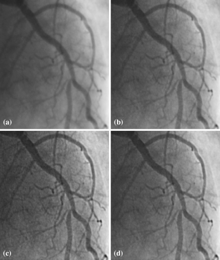
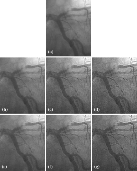
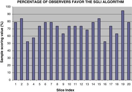
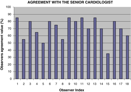
High quality visualization on X-ray angiograms is of great significance both for the diagnosis of vessel abnormalities and for coronary interventions. Algorithms for improving the visualization of detailed vascular structures without significantly increasing image noise are currently demanded in the market. A new algorithm called stick-guided lateral inhibition (SGLI) is presented for increasing the visibility of coronary vascular structures. A validation study was set up to compare the SGLI algorithm with the conventional unsharp masking (UM) algorithm on 20 still frames of coronary angiographic images. Ten experienced QCA analysts and nine cardiologists from various centers participated in the validation. Sample scoring value (SSV) and observer agreement value (OAV) were defined to evaluate the validation result, in terms of enhancing performance and observer agreement, respectively. The mean of SSV was concluded to be 77.1 +/- 11.9%, indicating that the SGLI algorithm performed significantly better than the UM algorithm (P-value < 0.001). The mean of the OAV was concluded to be 70.3%, indicating that the average agreement with respect to a senior cardiologist was 70.3%. In conclusion, this validation study clearly demonstrates the superiority of the SGLI algorithm in the visualization of coronary arteries from X-ray angiograms.
Figures

References
-
- None
- Reiber JHC, Koning G, Dijkstra J, Wahle A, Goedhart B, Sheehan FH, Sonka M (2000) Angiography and intravascular ultrasound. In: Sonka M, Fitzpatrick JM (eds) Handbook of medical imaging, vol 2. Medical image processing and analysis. SPIE, Washington, pp 711–808
-
- {'text': '', 'ref_index': 1, 'ids': [{'type': 'DOI', 'value': '10.1007/BF01145189', 'is_inner': False, 'url': 'https://doi.org/10.1007/bf01145189'}, {'type': 'PubMed', 'value': '8596059', 'is_inner': True, 'url': 'https://pubmed.ncbi.nlm.nih.gov/8596059/'}]}
- van der Zwet PMJ, Reiber JHC (1995) The influence of image enhancement and reconstruction on quantitative coronary arteriography. Int J Card Imaging 11:211–221 - PubMed
-
- {'text': '', 'ref_index': 1, 'ids': [{'type': 'DOI', 'value': '10.1117/1.482680', 'is_inner': False, 'url': 'https://doi.org/10.1117/1.482680'}]}
- Aach T, Schiebel U, Spekowius G (1999) Digital image acquisition and processing in medical X-ray imaging. J Electron Imaging 8(1):7–22
-
- Kumar MSD, Liyang W, Ram T, Jasjit SS (2007) DSA image enhancement via multi-resolution motion correction for interventional procedures: a robust strategy. Proceedings of the fifth IASTED international conference: biomedical engineering. ACTA Press, Innsbruck
-
- WIlson DL, Kaplan EJ (1990) Linear and morphological digital image enhancement of peripheral angiography images. Proceedings of SPIE: medical imaging IV: image processing. Newport Beach
Publication types
MeSH terms
LinkOut - more resources
Full Text Sources
Medical
