Review
. 2010 Jan;12(1):93-124.
doi: 10.1089/ars.2008.2425.

Redox control of asthma: molecular mechanisms and therapeutic opportunities

Affiliations
Review

Redox control of asthma: molecular mechanisms and therapeutic opportunities

Suzy A A Comhair et al. Antioxid Redox Signal. 2010 Jan.

Erratum in

  • Antioxid Redox Signal. 2010 Feb;12(2):321. Ghio,Andrew [removed]; Kinnula, Vuokko [removed]; Kliment, Corrine [removed];Montuschi, Paolo [removed]; Reddy, Sekhar [removed]; White, Carl [removed]

Abstract

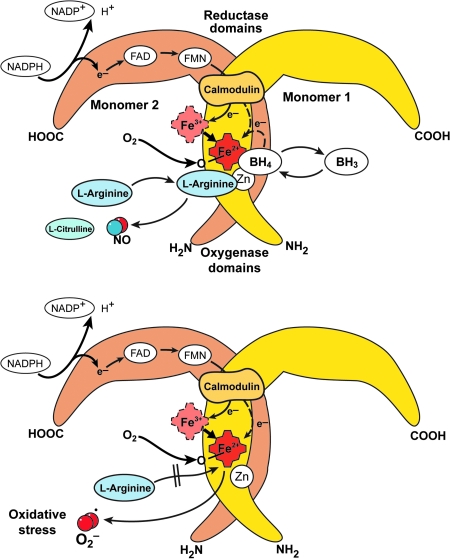
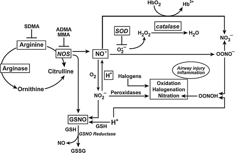
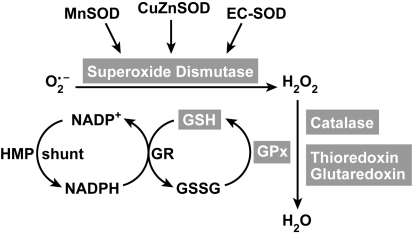
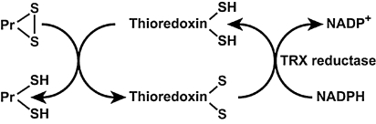
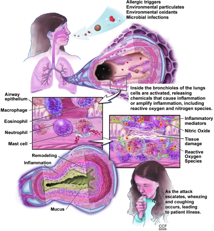
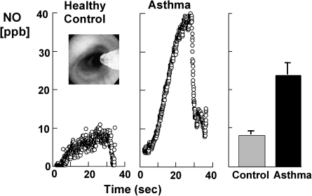
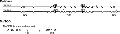
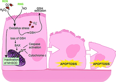
An imbalance in reducing and oxidizing (redox) systems favoring a more oxidative environment is present in asthma and linked to the pathophysiology of the defining symptoms and signs including airflow limitation, hyper-reactivity, and airway remodeling. High levels of hydrogen peroxide, nitric oxide ((*)NO), and 15-F(2t)-isoprostane in exhaled breath, and excessive oxidative protein products in lung epithelial lining fluid, peripheral blood, and urine provide abundant evidence for pathologic oxidizing processes in asthma. Parallel studies document loss of reducing potential by nonenzymatic and enzymatic antioxidants. The essential first line antioxidant enzymes superoxide dismutases (SOD) and catalase are reduced in asthma as compared to healthy individuals, with lowest levels in those patients with the most severe asthma. Loss of SOD and catalase activity is related to oxidative modifications of the enzymes, while other antioxidant gene polymorphisms are linked to susceptibility to develop asthma. Monitoring of exhaled (*)NO has entered clinical practice because it is useful to optimize asthma care, and a wide array of other biochemical oxidative and nitrative biomarkers are currently being evaluated for asthma monitoring and phenotyping. Novel therapeutic strategies that target correction of redox abnormalities show promise for the treatment of asthma.


Figures

FIG. 1.
FIG. 1.
Nicotinamide adenine dinucleotide (NAD+) functions in electron transfer reactions (redox) reactions. NAD+ acts as the oxidizing agent; it accepts electrons and become reduced to NADH. Subsequently, NADH serves as a reducing agent and donates electrons. Thus, NAD+ and NADH serve as a redox couple, as they accept and donate electrons in redox reactions, such as occur in cellular respiration. Multiple redox reactions constitute cellular respiration, in which oxygen is the terminal electron acceptor, and ATP is synthesized.
FIG. 2.
FIG. 2.
Sources of exogenous inhalational and endogenous reactive oxygen species (ROS) and reactive nitrogen species (RNS) in the lung. Environmental sources leading to greater amounts of ROS and RNS in the lungs are ozone, air pollutants (particulates as from diesel fuel combustion), particulates containing metals, and cigarette smoke. Endogenous ROS are produced as byproducts of mitochondrial respiration. Inflammatory cells can produce high levels of ROS and RNS in response to allergens and microbial infections. (For interpretation of the references to color in this figure legend, the reader is referred to the web version of this article at www.liebertonline.com/ars).
FIG. 3.
FIG. 3.
Production of reactive oxygen species (ROS). Superoxide (O2) reacts rapidly with itself, or is catalytically converted by superoxide dismutases (SOD), to form hydrogen peroxide (H2O2). Hydrogen peroxide is detoxified to water by catalase or glutathione peroxidase enzymes (GPx). Extremely toxic reactions of superoxide and hydrogen peroxide that form hydrogen radical occur via the Haber–Weiss and Fenton chemistry reactions in the presence of metal ions. Hydrogen peroxide is converted by myeloperoxide (MPO) or eosinophil peroxidase (EPO) to highly reactive halogenating acids, such as hypobromous acid (HOBr) or hypochlorous acid (HOCl), xanthine oxidase (XO). (For interpretation of the references to color in this figure legend, the reader is referred to the web version of this article at www.liebertonline.com/ars).
FIG. 4.
FIG. 4.
Amino acid oxidation products and cross-links formed by peroxidase enzymes. Protein oxidative damage mediated by EPO-generated reactive brominating species (RBS), MPO-generated reactive chlorinating species (RCS), reactive nitrating species (RNS), tyrosyl radical (oY), transition metal ions (M2+) that form hydroxyl radical, may be identified by stable products formed by each pathway. All are tyrosine derivatives, except the nonphysiologic o-tyrosine and m-tyrosine that form from the oxidation of phenylalanine.
FIG. 5.
FIG. 5.
Nitric oxide synthases (NOS). General schematic for all active NOS shows that the dimeric enzymes are comprised of two monomers. The oxygenase domains of two subunits interact to form the homodimer. NOS convert L-arginine to NO and L-citrulline in a reaction that requires oxygen, NADPH, and cofactors FAD, FMN, tetrahydrobiopterin, calmodulin, and iron protoporphyrin IX. The N-terminus oxygenase domain of each monomer binds the heme, tetrahydrobiopterin, and substrate L-arginine. The carboxy terminus of each monomer binds the FAD, FMN, and NADPH. The carboxy-reductase domain of one monomer transfers electrons from NADPH to FAD to FMN and ultimately to the oxygenase domain ferric heme iron of the other monomer, which then binds oxygen and oxidizes L-arginine to generate NO and citrulline. NO synthesis is regulated by availability of substrate L-arginine and cofactor tetrahydrobiopterin. In ‘coupled’ NOS, tetrahydrobiopterin enables electrons from NADPH to be used for NO synthesis. In ‘uncoupled’ NOS, oxygen reduction occurs but results in superoxide or H202 release instead of NO.
FIG. 6.
FIG. 6.
Redox chemistry in the lung. Levels of NO and other nitrogen oxides, superoxide, and other reactive oxygen species, are regulated both enzymatically and by nonenzymatic reactions. Arginase enzymes serve as a metabolic branch point controlling the flow of L-arginine to protein synthesis, NO synthesis, and ornithine and urea cycle. Arginase activity is increased in asthmatic lungs. Ornithine is a precursor for polyamines and proline for cell proliferation and collagen synthesis, respectively, critical components of airway remodeling. Once formed, NO may react rapidly with O2•− yielding ONOO-. Following ONOO- protonation, ONOOH can nitrate tyrosine (Tyr-NO2) or convert to NO3-. NO2- formation from NO is slow. Rather, NO2 protonation to form NO is favored in the increased acidity that is present in the asthmatic airway. NO3 is present at higher than normal levels in the oxidizing acidic environment of the asthmatic lung, but NO2*#x2212; is similar in asthmatic and control lungs. NO2- is also consumed in leukocyte peroxidase (EPO and MPO) catalyzed reactions, which also generate halogenating reactive species. Nitrosoglutathione (GSNO) is a beneficial endogenous bronchodilator that is catabolized by GSNO reductase to release NO.
FIG. 7.
FIG. 7.
Antioxidants in redox reactions. Superoxide can be detoxified by superoxide dismutases (SOD). There are three forms: an intracellular CuZnSOD, mitochondrial MnSOD, and an extracellular EC-SOD. Hydrogen peroxide (H202) can be further detoxified to water by catalase, thioredoxin (TRX), glutoredoxin (GRX) and/or by the glutathione peroxidase (GPx). TRX, GRX and GPx use glutathione as a cofactor. The oxidized glutathione (GSSG) is subsequently returned to GSH by glutathione reductase, an intracellular enzyme that uses NADPH generated from the hexose monophosphate shunt system (HMP shunt) as an electron donor.
FIG. 8.
FIG. 8.
Thioredoxin redox system. Thioredoxins [Thioredoxin-(SH)2] act as proton donors and cleave disulfide (S–S) bonds in target proteins [P-(S–S)]. Thioredoxin reductase is responsible for reconstitution of the reduced thioredoxin from the oxidized form [thioredoxin-(S–S)].
FIG. 9.
FIG. 9.
Glutaredoxin system. Glutaredoxins (GRX) are thiol–disulfide oxidoreductases that catalyze the reversible exchange of GSH with protein thiol groups (PrSH). Dithiol GRXs contain Cys-Pro-Tyr-Cys active site motif and monothiol GRXs have Cys-Gly-Phe-Ser active sites. Modifed from Hurd et al. (150).
FIG. 10.
FIG. 10.
Pathophysiology of the inflammation and redox abnormalities in asthma. 137 × 177 mm (300 × 300 DPI).
FIG. 11.
FIG. 11.
Increased Inflammatory cells in asthmatic airways. Immunohistochemistry of endobronchial biopsies obtained from asthmatic lungs show the presence of increased numbers of polymorphonuclear cells (A, B), eosinophils (C, D) and mast cells (E, F) infiltrating throughout the mucosa and submucosa. Other remodeling changes seen in the biopsies include thickened basement membrane and sloughing of the surface epithelium (seen in C), increased vascularity (identifiable in A), and hypertrophy of the smooth muscle cells and layer (seen in C and E).
FIG. 12.
FIG. 12.
High levels of nitric oxide production and nitrotyrosine in asthma. Kinetics of NO accumulation in the gas phase in the airway lumen (left panel) are shown over time of a breath hold of a healthy control and an asthmatic individual. Individuals underwent bronchoscopy with a flexible fiberoptic bronchoscope and the levels of NO measured at a segmental bronchus with a collection Teflon catheter adapted to the working channel of the flexible bronchoscope (inset picture shows the catheter in the airway lumen). Sampling is performed in bronchioles between 5–7 mm in diameter. Individuals perform breath-hold (20 sec) and the accumulation of NO recorded in the absence of airflow. This type of evaluation yields a plot of NO (ppb) versus time (sec). During the breath-hold, bronchiolar gases accumulate NO quickly to a plateau. At the end of expiratory breath-hold, individuals exhale completely and this is accompanied by a rapid drop of NO as alveolar gases, which do not accumulate NO, are delivered to the sampling catheter. Levels of NO are measured with chemiluminescent analyser (NOA 280 Sievers) adapted for on-line data recording of NO concentration [methods as in Dweik et al. (90)]. Asthmatics generate levels of NO in the airway that are higher than healthy controls. Nitrotyrosine immunostaining of asthmatic and healthy control bronchial mucosa is shown in the right panel. Healthy control bronchial mucosa has pseudostratified columnar epithelium, with nitrotyrosine (red) staining present in apical portions of cells. Asthmatic bronchial mucosa has marked increase in immunoreactivity for nitrotyrosine in the epithelial cells. There are increased numbers of goblet cells in the biopsy, which are seen as cells with clear, nonstaining intracellular areas. Figures are modified from Dweik et al. (92).
FIG. 13.
FIG. 13.
Tyrosine in catalase and MnSOD. Sequence location of 20 tyrosines in catalase and 10 tyrosine in MnSOD. (*) indicates sequence difference between murine and human. In catalase, Tyr 358 (filled circle) binds the proximal heme ligand and is critical for enzyme activity. Catalase contains a putative chlororination site (KXHY) at Tyr236. MnSOD Tyr34 (filled circle) is located in the active site of the enzyme and modification leads to inactivation of the enzyme. Figures are modified from Ghosh et al. (119).
FIG. 14.
FIG. 14.
Redox abnormalities trigger apoptosis in airway epithelial cells. Exposure to ROS and/or RNS leads to extrusion of intracellular GSH and GSSG, and oxidative modification of MnSOD. Loss of SOD activity and/or extrusion of GSH activates BAX and caspases, and causes cytochrome c release from mitochondria, all of which trigger cell entry into programmed cell death pathways. This mechanism likely contributes to apoptosis and loss of airway epithelial cells, which is a hallmark of the remodeling in the asthmatic airway. (For interpretation of the references to color in this figure legend, the reader is referred to the web version of this article at www.liebertonline.com/ars).

References

    1. Abu-Soud HM. Hazen SL. Nitric oxide is a physiological substrate for mammalian peroxidases. J Biol Chem. 2000;275:37524–37532. - PubMed
    1. Abu-Soud HM. Hazen SL. Nitric oxide modulates the catalytic activity of myeloperoxidase. J Biol Chem. 2000;275:5425–5430. - PubMed
    1. Abu-Soud HM. Khassawneh MY. Sohn JT. Murray P. Haxhiu MA. Hazen SL. Peroxidases inhibit nitric oxide (NO) dependent bronchodilation: Development of a model describing NO-peroxidase interactions. Biochemistry. 2001;40:11866–11875. - PubMed
    1. Adcock IM. Brown CR. Kwon O. Barnes PJ. Oxidative stress induces NF kappa B DNA binding and inducible NOS mRNA in human epithelial cells. Biochem Biophys Res Commun. 1994;199:1518–1524. - PubMed
    1. Agani FH. Pichiule P. Chavez JC. LaManna JC. The role of mitochondria in the regulation of hypoxia-inducible factor 1 expression during hypoxia. J Biol Chem. 2000;275:35863–35867. - PubMed

Publication types