. 2009 Jul 27:7:44.
doi: 10.1186/1741-7007-7-44.

A Dictyostelium chalone uses G proteins to regulate proliferation

Affiliations

A Dictyostelium chalone uses G proteins to regulate proliferation

Deenadayalan Bakthavatsalam et al. BMC Biol. .

Abstract

Background: Several studies have shown that organ size, and the proliferation of tumor metastases, may be regulated by negative feedback loops in which autocrine secreted factors called chalones inhibit proliferation. However, very little is known about chalones, and how cells sense them. We previously identified two secreted proteins, AprA and CfaD, which act as chalones in Dictyostelium. Cells lacking AprA or CfaD proliferate faster than wild-type cells, and adding recombinant AprA or CfaD to cells slows their proliferation.

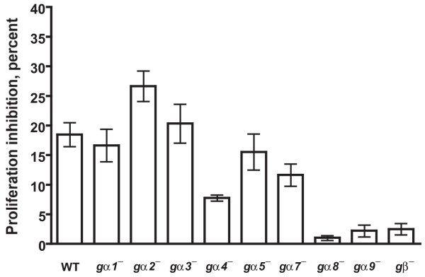
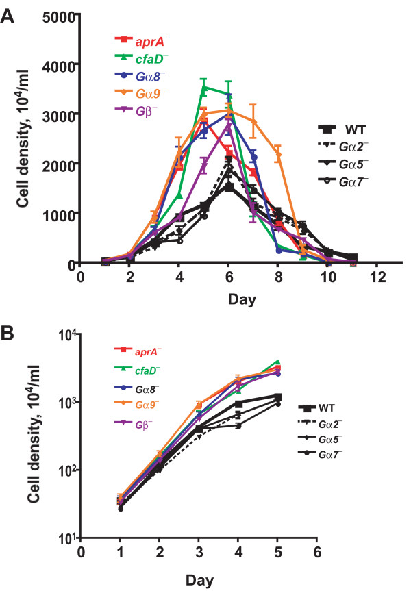
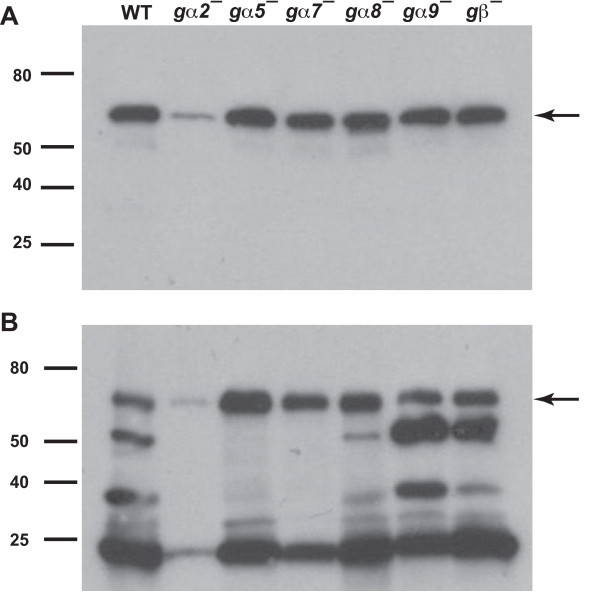
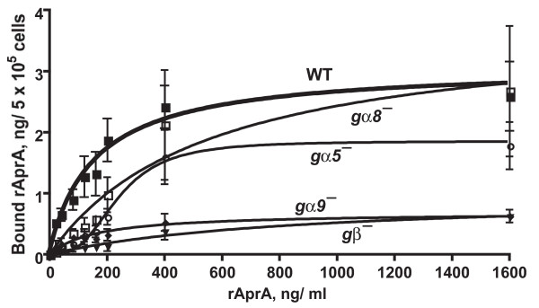
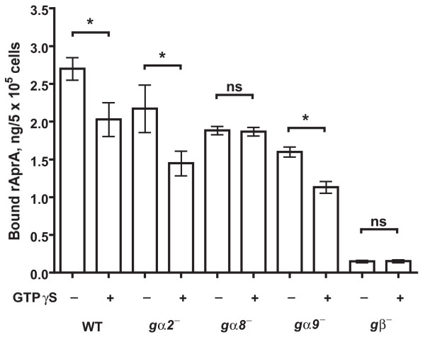
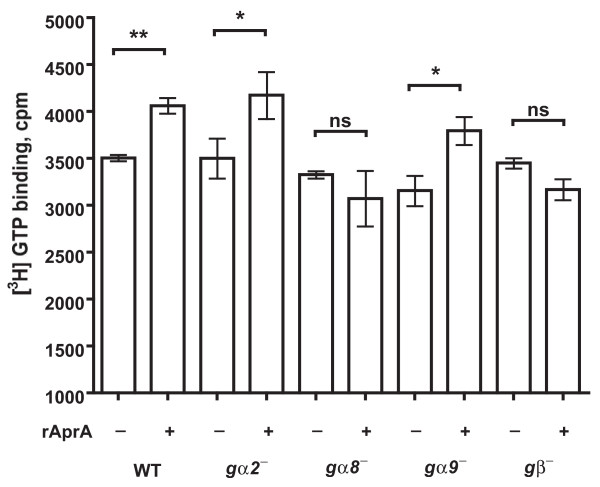
Results: We show here that cells lacking the G protein components Galpha8, Galpha9, and Gbeta proliferate faster than wild-type cells despite secreting normal or high levels of AprA and CfaD. Compared with wild-type cells, the proliferation of galpha8-, galpha9- and gbeta- cells are only weakly inhibited by recombinant AprA (rAprA). Like AprA and CfaD, Galpha8 and Gbeta inhibit cell proliferation but not cell growth (the rate of increase in mass and protein per nucleus), whereas Galpha9 inhibits both proliferation and growth. galpha8- cells show normal cell-surface binding of rAprA, whereas galpha9- and gbeta- cells have fewer cell-surface rAprA binding sites, suggesting that Galpha9 and Gbeta regulate the synthesis or processing of the AprA receptor. Like other ligands that activate G proteins, rAprA induces the binding of [3H]GTP to membranes, and GTPgammaS inhibits the binding of rAprA to membranes. Both AprA-induced [3H]GTP binding and the GTPgammaS inhibition of rAprA binding require Galpha8 and Gbeta but not Galpha9. Like aprA- cells, galpha8- cells have reduced spore viability.

Conclusion: This study shows that Galpha8 and Gbeta are part of the signal transduction pathway used by AprA to inhibit proliferation but not growth in Dictyostelium, whereas Galpha9 is part of a differealnt pathway that regulates both proliferation and growth, and that a chalone signal transduction pathway uses G proteins.


Figures

Figure 1
Figure 1
The effect of recombinant AprA (rAprA) on cell proliferation. Cells were grown in the presence or absence of rAprA for 12 h, and the percent decrease in cell density caused by rAprA was calculated. When compared with the inhibition of wild-type (WT) cell proliferation, the inhibition of gα1- , gα2-, gα3-, gα5-, and gα7- cells is not significant with P > 0.05 (one-way ANOVA, Dunnett's test), whereas the inhibition of gα4- is significant with P < 0.01, and the inhibition of gα8-, gα9-, and - cells is significant with P < 0.001. The inhibition values for gα8-, gα9-, and - cells are not significantly different between each other with P > 0.05 (one-way ANOVA, Tukey's test), but the inhibition of Gα4- cells is significantly different from that of gα8-, gα9-, and - cells with P < 0.01 (one-way ANOVA, Dunnett's test). Values are the mean ± s.e.m. from at least four separate experiments.
Figure 2
Figure 2
Cells lacking Gα8, Gα9 or Gβ proliferate faster than wild-type cells. (A) Cells were diluted to 2 × 10⁵cells/ml in HL5 and the cell density was measured daily. The graph shows means ± s.e.m. from three independent experiments. The differences between the maximum cell density attained by wild-type cells and aprA-, cfaD-, gα8-, gα9-, and - mutants are significant with P < 0.05, whereas the difference between the wild-type maximum density and the gα2-, gα5-, and gα7- maximum densities are not significant (one-way ANOVA, Dunnett's test). (B) The data from the first five days was plotted using a log scale for the density. The absence of error bars indicates that the error was smaller than the plot symbol.
Figure 3
Figure 3
The accumulation of extracellular AprA and CfaD. The indicated cell types were grown in HL5 and conditioned growth media were collected. Western blots of the conditioned growth media were stained with anti-AprA antibodies (A) or anti-CfaD antibodies (B). The arrow in A indicates the 60 kDa AprA band, and the arrow in B indicates the 62 kDa CfaD band. The lower molecular mass bands stained in B are breakdown products of CfaD [3]. Data are representative of three independent experiments.
Figure 4
Figure 4
The binding of recombinant AprA to cells. The indicated cells (WT is wild-type) were incubated with different concentration of recombinant AprA (rAprA). After 10 min, bound rAprA was quantitated by Western blots (staining for the Myc tag), using known amounts of rAprA as standards. Values are mean ± s.e.m. (n = 3). The lines are curve fits to a one-site binding model with no cooperative binding with the exception of the fit to the binding to gα5- cells, where the line is a fit to a one-site binding model with a Hill coefficient of 2.9.
Figure 5
Figure 5
The effect of GTPγS on recombinant AprA binding to membranes. Membranes from the indicated strains were incubated with recombinant AprA (rAprA) in the presence or absence of GTPγS. rAprA bound to the membranes was measured as in Figure 4. The presence of GTPγS significantly decreased the binding of rAprA to WT, gα2- and gα9- membranes in comparison with the buffer control (*, P < 0.05), while the presence of GTPγS had no significant effect (ns) on the binding of rAprA to gα8- and - membranes (paired t-tests). Values are mean ± s.e.m. (n = 3).
Figure 6
Figure 6
The effect of recombinant AprA on GTP binding to membranes. The binding of [3H]GTP to membranes in the presence or absence of recombinant AprA (rAprA) was measured following [27]. Wild-type membranes showed significantly increased [3H]GTP binding in the presence of rAprA when compared with [3H]GTP binding in the absence of rAprA with P < 0.01 (**), while gα2- and gα9- membranes showed increased [3H]GTP binding with P < 0.05 (*). [3H]GTP binding to gα8- and - membranes was not significantly different (ns) (paired t-tests). Values are the mean ± s.e.m of at least three independent experiments.

Similar articles

Cited by

References

    1. Twitty VC, Schwind JL. The growth of eyes and limbs transplanted heteroplastically between two species of Amblystomt. J Exp Zool. 1931;59:61–86. doi: 10.1002/jez.1400590105. - DOI
    1. Elgjo K, Reichelt KL. Chalones: from aqueous extracts to oligopeptides. Cell Cycle. 2004;3:1208–1211. - PubMed
    1. Bakthavatsalam D, Brock DA, Nikravan NN, Houston KD, Hatton RD, Gomer RH. The secreted Dictyostelium protein CfaD is a chalone. J Cell Sci. 2008;121:2473–2480. doi: 10.1242/jcs.026682. - DOI - PMC - PubMed
    1. Brock DA, Gomer RH. A secreted factor represses cell proliferation in Dictyostelium. Development. 2005;132:4553–4562. doi: 10.1242/dev.02032. - DOI - PMC - PubMed
    1. Lee S, McPherron A. Myostatin and the control of skeletal muscle mass. Curr Opin Genet Dev. 1999;9:604–607. doi: 10.1016/S0959-437X(99)00004-0. - DOI - PubMed

Publication types

MeSH terms

LinkOut - more resources