. 2009 Jul 27:5:26.
doi: 10.1186/1746-6148-5-26.

Immunohistochemical and biochemical characteristics of BSE and CWD in experimentally infected European red deer (Cervus elaphus elaphus)

Affiliations

Immunohistochemical and biochemical characteristics of BSE and CWD in experimentally infected European red deer (Cervus elaphus elaphus)

Stuart Martin et al. BMC Vet Res. .

Abstract

Background: The cause of the bovine spongiform encephalopathy (BSE) epidemic in the United Kingdom (UK) was the inclusion of contaminated meat and bone meal in the protein rations fed to cattle. Those rations were not restricted to cattle but were also fed to other livestock including farmed and free living deer. Although there are no reported cases to date of natural BSE in European deer, BSE has been shown to be naturally or experimentally transmissible to a wide range of different ungulate species. Moreover, several species of North America's cervids are highly susceptible to chronic wasting disease (CWD), a transmissible spongiform encephalopathy (TSE) that has become endemic. Should BSE infection have been introduced into the UK deer population, the CWD precedent could suggest that there is a danger for spread and maintenance of the disease in both free living and captive UK deer populations. This study compares the immunohistochemical and biochemical characteristics of BSE and CWD in experimentally-infected European red deer (Cervus elpahus elaphus).

Results: After intracerebral or alimentary challenge, BSE in red deer more closely resembled natural infection in cattle rather than experimental BSE in small ruminants, due to the lack of accumulation of abnormal PrP in lymphoid tissues. In this respect it was different from CWD, and although the neuropathological features of both diseases were similar, BSE could be clearly differentiated from CWD by immunohistochemical and Western blotting methods currently in routine use.

Conclusion: Red deer are susceptible to both BSE and CWD infection, but the resulting disease phenotypes are distinct and clearly distinguishable.


Figures

Figure 1
Figure 1
BSE deer. Vacuolation seen in the brainstem (a), the molecular layer of the cerebellum (b), the thalamus (c), and layers V and VI of the cerebral cortex (d).
Figure 2
Figure 2
BSE deer. Grey matter PrPd labelling of neuropil and intense perikaryonal labelling of a Golgi neuron in the granular cell layer of the cerebellum (arrow).
Figure 3
Figure 3
CWD Deer. Vacuolation (oval) and diffuse PrPd accumulation is present in the grey matter of the striatum of the brain. Focal intense plaque-like accumulations of PrPd (boxes) are also present.
Figure 4
Figure 4
IHC profile of PrPd types present in BSE infected deer (green) and CWD infected deer (red). Profiles represent a single deer from each of the BSE and CWD infected groups. All six of the deer challenged i.c. with BSE and the single positive orally dosed deer presented consistent profiles on subjective examination that were unlike those of the CWD infected deer. (ITNR, intraneuronal; ITAS, intra-astrocytic; ITMG, intra-microglial; STEL, stellate; SBPL, sub-pial; SBEP, sub-ependymal; PVRS, peri-vascular; PVAC, peri-vacuolar; PRCO, particulate coalescing; LINR, linear; PNER, perineuronal; VASC, vascular; EPEN, Ependymal).
Figure 5
Figure 5
BSE and CWD deer. Low magnification images (top) of midbrain highlight the marked differences seen between the predominantly intra-neronal and difuse particulate labelling of the neuropil that is seen in the BSE infected deer compared to the distinct coalescing plaque-like labelling in the CWD infected deer. The boxes outline the area shown at higher magnification in the lower two images.
Figure 6
Figure 6
BSE deer. Comparison of intraneuronal labelling with BAR224 (C-terminal) and 12B2 (n-terminal) mAbs showing the significant loss of intraneuronal labelling with 12B2 in BSE infected deer in contrast to CWD infected deer. The extracellular signal was maintained with 12B2 in both the CWD and BSE infected deer.
Figure 7
Figure 7
CWD deer. Comparison of intraneuronal labelling with BAR224 (C-terminal) and 12B2 (n-terminal) mAbs showing no reduction of the intracellular signal with the n-terminal targeting antibody.
Figure 8
Figure 8
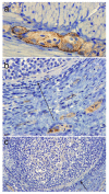
BSE deer. Labelling of PrPd in cells of the myenteric (a) and sub-mucosal plexi (b) occurred in close proximity to negative Peyer's patches shown at high (b) and low magnification (c). Arrows show the capsule surrounding the same follicle.
Figure 9
Figure 9
CWD Deer. Labelling of PrPd in tingible body macrophages (a) and follicular dendritic cells (b) within the secondary follicle of a retropharyngeal lymph node.
Figure 10
Figure 10
BSE deer. Analysis of brain homogenates by Biorad TeSeE Western Blotting using F99, 6H4, SHA31, P4 and 12B2. Data is not shown for the last of the intracerebrally challenged group or for the single orally challenged BSE positive deer however these presented migration patterns and antibody affinities that were not significantly different from those seen above.
Figure 11
Figure 11
CWD deer. Analysis of brain homogenates by Biorad TeSeE Western Blotting using F99, 6H4, SHA31, P4 and 12B2. The molecular weights of samples from the four red deer experimentally infected with CWD were variable although all of them appeared higher than the cattle BSE control and are more consistent with the naturally infected CWD and scrapie controls.

References

    1. Wilesmith JW, Wells GAH, Cranwell MP, Ryan JBN. BSE: Epidemiological Studies. Vet Record. 1988;123:638–644. - PubMed
    1. Jeffrey M, Wells GA. Spongiform encephalopathy in a nyala (Tragelaphus angasi) Vet Pathol. 1988;25:398–399. - PubMed
    1. Seuberlich T, Botteron C, Wenker C, Cafe-Marcal V, Oevermann A, Haase B, et al. Spongiform encephalopathy in a miniature zebu. Emerg Infect Dis. 2006;12:1950–1953. - PMC - PubMed
    1. Kirkwood JK, Cunningham AA. Epidemiologic Observations on Spongiform Encephalopathies in Captive Wild Animals in the British-Isles. Veterinary Record. 1994;135:296–303. - PubMed
    1. Williams ES. Chronic wasting disease. Vet Pathol. 2005;42:530–549. doi: 10.1354/vp.42-5-530. - DOI - PubMed

Publication types