. 2009 Jul 28:9:44.
doi: 10.1186/1471-213X-9-44.

Nucleologenesis and embryonic genome activation are defective in interspecies cloned embryos between bovine ooplasm and rhesus monkey somatic cells

Affiliations

Nucleologenesis and embryonic genome activation are defective in interspecies cloned embryos between bovine ooplasm and rhesus monkey somatic cells

Bong-Seok Song et al. BMC Dev Biol. .

Abstract

Background: Interspecies somatic cell nuclear transfer (iSCNT) has been proposed as a tool to address basic developmental questions and to improve the feasibility of cell therapy. However, the low efficiency of iSCNT embryonic development is a crucial problem when compared to in vitro fertilization (IVF) and intraspecies SCNT. Thus, we examined the effect of donor cell species on the early development of SCNT embryos after reconstruction with bovine ooplasm.

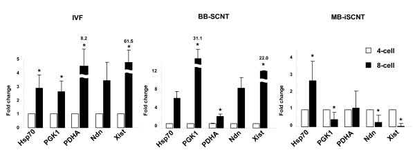
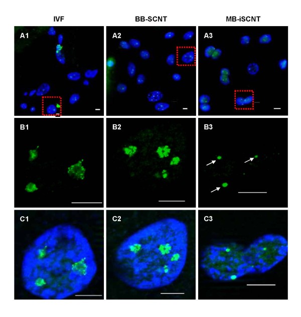
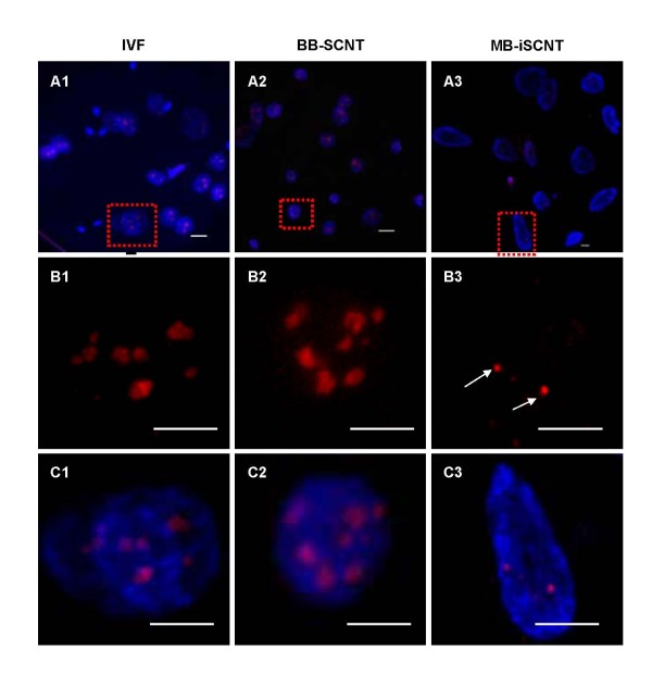
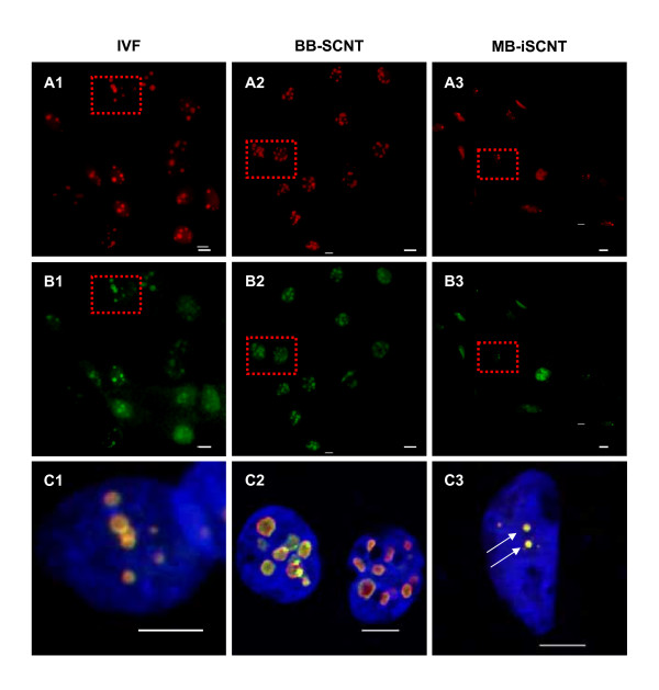
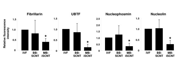
Results: No apparent difference in cleavage rate was found among IVF, monkey-bovine (MB)-iSCNT, and bovine-bovine (BB)-SCNT embryos. However, MB-iSCNT embryos failed to develop beyond the 8- or 16-cell stages and lacked expression of the genes involved in embryonic genome activation (EGA) at the 8-cell stage. From ultrastructural observations made during the peri-EGA period using transmission electron microscopy (TEM), we found that the nucleoli of MB-iSCNT embryos were morphologically abnormal or arrested at the primary stage of nucleologenesis. Consistent with the TEM analysis, nucleolar component proteins, such as upstream binding transcription factor, fibrillarin, nucleolin, and nucleophosmin, showed decreased expression and were structurally disorganized in MB-iSCNT embryos compared to IVF and BB-SCNT embryos, as revealed by real-time PCR and immunofluorescence confocal laser scanning microscopy, respectively.

Conclusion: The down-regulation of housekeeping and imprinting genes, abnormal nucleolar morphology, and aberrant patterns of nucleolar proteins during EGA resulted in developmental failure in MB-iSCNT embryos. These results provide insight into the unresolved problems of early embryonic development in iSCNT embryos.


Figures

Figure 1
Figure 1
Analysis of mitochondrial DNA (mtDNA) in monkey-bovine MB-iSCNT embryos on day 3. Rhesus monkey ear skin fibroblasts (A), image of MB-iSCNT embryos at day 3 (B), and expression of mtDNA in MB-iSCNT embryos. Bar, 50 μm. B, bovine; M, rhesus monkey.
Figure 2
Figure 2
Real-time PCR quantification of mRNA expression in IVF, BB-SCNT, and MB-iSCNT embryos. Relative abundance of housekeeping and imprinting genes in IVF, BB-SCNT, and MB-iSCNT embryos between the 4- (before EGA) and 8-cell stages (after EGA). *Values are significantly different from the 4-cell control (P < 0.05).
Figure 3
Figure 3
TEM evaluation of nucleolar developmental stages in IVF, BB-SCNT, and MB-iSCNT embryos. Primary vacuole (a1, white arrow) and both primary and secondary vacuoles (a2, white arrow head) in an IVF embryo. Both primary and secondary vacuoles (b1) and reticular nucleolus (b2, black arrow) in a BB-SCNT embryo. NPBs (c1, black arrow head) and abnormally structured nucleolus (c2, gray arrows) in an MB-iSCNT embryo. (B) Proportion of each nucleolar developmental stage in IVF, BB-SCNT, and MB-iSCNT embryos. (C) Proportion of each nucleolar development stage in both 8-cell and 16-cell stage embryos. Bar, 2 μm (A1, A2, B1, and C1); 1 μm (B2 and C2).
Figure 4
Figure 4
Expression and localization of upstream binding transcription factor (UBTF) during embryonic genome activation (EGA). IVF (A1, B1, and C1) and BB-SCNT (A2, B2, and C2) embryos showed clusters of foci, whereas MB-iSCNT embryos showed only small foci (A3, B3, and C3). DNA was stained with DAPI. Arrow indicates small UBTF foci in an MB-iSCNT embryo. Bar, 10 μm.
Figure 5
Figure 5
Expression and localization of fibrillarin proteins during embryonic genome activation (EGA). IVF (A1, B1, and C1), BB-SCNT (A2, B2, and C2), and MB-iSCNT (A3, B3, and C3) embryos were immunostained with antibodies specific to fibrillarin. DNA was stained with DAPI. Arrow indicates small fibrillarin foci in an MB-iSCNT embryo. Bar, 10 μm.
Figure 6
Figure 6
Expression and localization of the nucleophosmin and nucleolin proteins during embryonic genome activation (EGA). IVF (A1, B1, and C1), BB-SCNT (A2, B2, and C2), and MB-iSCNT (A3, B3, and C3) embryos were immunostained with antibodies specific to nucleophosmin and nucleolin. DNA was stained with DAPI. Arrow indicates small foci of nucleophosmin and nucleolin staining in an MB-iSCNT embryo. Bar, 10 μm.
Figure 7
Figure 7
Evaluation of nucleolar protein expression in embryos derived from IVF, BB-SCNT, and MB-iSCNT. The expression of UBTF, fibrillarin, nucleophosmin, and nucleolin were relative to the mean expressed cells to the total nuclei in early (A) and late (B) EGA-stage embryos. Early EGA, 8- to 11-cell stage; late EGA, 12- to 16-cell stage. *Values are significantly different from the other groups (P < 0.05).

References

    1. Illmensee K, Levanduski M, Zavos PM. Evaluation of the embryonic preimplantation potential of human adult somatic cells via an embryo interspecies bioassay using bovine oocytes. Fertil Steril. 2006;85:1248–60. doi: 10.1016/j.fertnstert.2005.10.026. - DOI - PubMed
    1. Chen T, Zhang YL, Jiang Y, Liu JH, Schatten H, Chen DY, Sun QY. Interspecies nuclear transfer reveals that demethylation of specific repetitive sequences is determined by recipient ooplasm but not by donor intrinsic property in cloned embryos. Mol Reprod Dev. 2006;73:313–7. doi: 10.1002/mrd.20421. - DOI - PubMed
    1. Chang KH, Lim JM, Kang SK, Lee BC, Moon SY, Hwang WS. An optimized protocol of a human-to-cattle interspecies somatic cell nuclear transfer. Fertil Steril. 2004;82:960–2. doi: 10.1016/j.fertnstert.2004.03.052. - DOI - PubMed
    1. Zhao ZJ, Ouyang YC, Nan CL, Lei ZL, Song XF, Sun QY, Chen DY. Rabbit oocyte cytoplasm supports development of nuclear transfer embryos derived from the somatic cells of the camel and Tibetan antelope. J Reprod Dev. 2006;52:449–59. doi: 10.1262/jrd.17095. - DOI - PubMed
    1. Arat S, Rzucidlo SJ, Stice SL. Gene expression and in vitro development of inter-species nuclear transfer embryos. Mol Reprod Dev. 2003;66:334–42. doi: 10.1002/mrd.10362. - DOI - PubMed

Publication types

LinkOut - more resources