. 2009 Nov;89(5):774-81.
doi: 10.1016/j.exer.2009.07.010. Epub 2009 Jul 25.

Vascular adhesion protein-1 regulates leukocyte transmigration rate in the retina during diabetes

Affiliations

Vascular adhesion protein-1 regulates leukocyte transmigration rate in the retina during diabetes

Kousuke Noda et al. Exp Eye Res. 2009 Nov.

Abstract

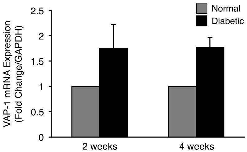
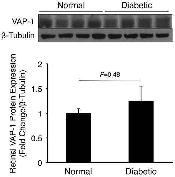
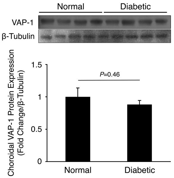
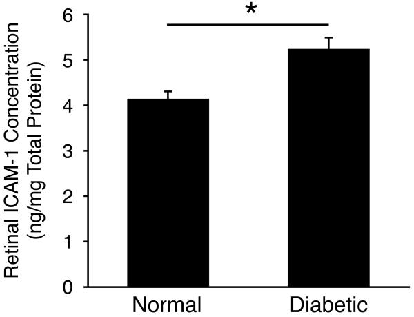
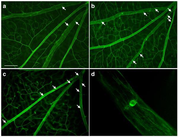
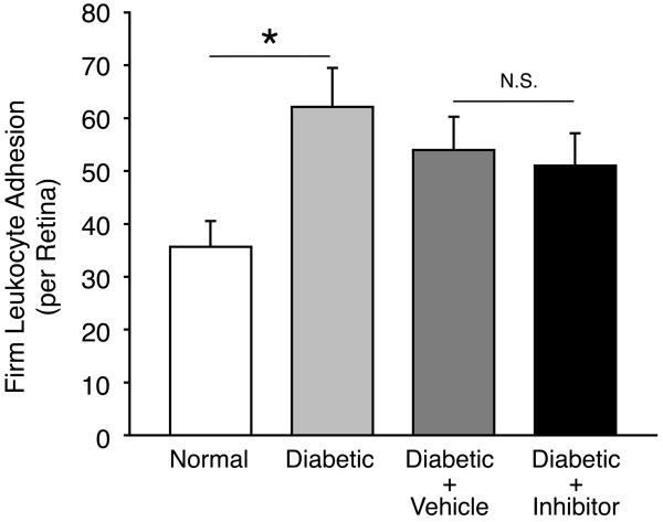
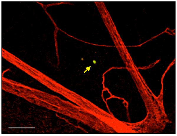
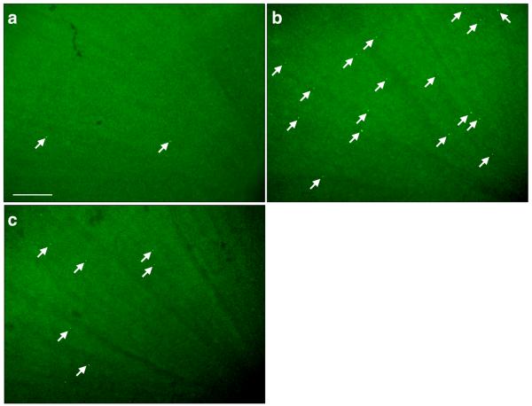
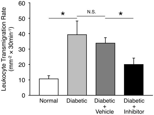
Vascular adhesion protein-1 (VAP-1) is an endothelial adhesion molecule that possesses semicarbazide-sensitive amine oxidase (SSAO) activity and is involved in leukocyte recruitment. Leukocyte adhesion to retinal vessels is a predominant feature of experimentally induced diabetic retinopathy (DR). However, the role of VAP-1 in this process is unknown. Diabetes was induced by i.p. injection of Streptozotocin in Long-Evans rats. The specific inhibitor of VAP-1, UV-002, was administered by daily i.p. injections. The expression of VAP-1 mRNA in the retinal extracts of normal and diabetic animals was measured by real-time quantitative polymerase chain reaction (PCR). Firm leukocyte adhesion was quantified in retinal flatmounts after intravascular staining with concanavalin A (ConA). Leukocyte transmigration rate was quantified by in vivo acridine orange leukocyte staining (AOLS). In diabetic rats, the rate of leukocyte transmigration into the retinal tissues of live animals was significantly increased, as determined by AOLS. When diabetic animals were treated with daily injections of the VAP-1 inhibitor (0.3 mg/kg), leukocyte transmigration rate was significantly reduced (P < 0.05). However, firm adhesion of leukocytes in diabetic animals treated with the inhibitor did not differ significantly from vehicle-treated diabetic controls. This work provides evidence for an important role of VAP-1 in the recruitment of leukocyte to the retina in experimental DR. Our results reveal the critical contribution of VAP-1 to leukocyte transmigration, with little impact on firm leukocyte adhesion in the retinas of diabetic animals. VAP-1 inhibition might be beneficial in the treatment of DR.


Figures

Figure 1
Figure 1. Retinal VAP-1 mRNA Expression in Normal and Diabetic Animals
Quantitative real-time PCR analysis showing increased retinal VAP-1 expression in diabetic animals compared to normal controls. Values are mean±SEM (n=11 and 12 in each group, 2 and 4 weeks after diabetes induction, respectively). * P<0.05.
Figure 2
Figure 2. Retinal and Choroidal VAP-1 Expression in Normal and Diabetic Animals
Western blot analysis of VAP-1 and β-tubulin expression in (A) retinal and (B) choroidal samples of normal and 2 weeks diabetic animals (n=4 in each group).
Figure 2
Figure 2. Retinal and Choroidal VAP-1 Expression in Normal and Diabetic Animals
Western blot analysis of VAP-1 and β-tubulin expression in (A) retinal and (B) choroidal samples of normal and 2 weeks diabetic animals (n=4 in each group).
Figure 3
Figure 3. Retinal ICAM-1 Expression in Diabetes
ICAM-1 protein expression in retinal tissues of normal and diabetic animals (n=4 in each group).
Figure 4
Figure 4. Contribution of VAP-1 to Firm Leukocyte Adhesion in the Retinal Vessels During Diabetes
(A) Representative micrographs of the ConA-stained flat-mounted retinas obtained from control and diabetic animals with or without VAP-1 inhibitor treatment. a) Normal control, b) Diabetic + vehicle, c) Diabetic + VAP-1 inhibitor, and d) Magnified view of a firmly adhering leukocyte in the retinal vein of a diabetic animal. Arrows depict firmly adhering leukocytes in the retinal vessels. Bar, 100μm. (B) Bars represent the average numbers of firmly adhering leukocytes in retinal vessels of normal and diabetic animals, treated with VAP-1 inhibitor or control from ConA-stained whole retinal flat-mounts. Error bars represent SEM (n=6 in each group), * P<0.05; N.S., not significant.
Figure 5
Figure 5. Impact of VAP-1 Inhibition on the Retinal Leukocyte Extravasation Rate During Diabetes
(A) Three dimensional reconstruction of confocal sections from the retina of a diabetic animal, 30 minutes after systemic AO injection and cardiac perfusion with rhodamine conjugated ConA. Arrow indicates an AO-stained leukocyte transmigrated out of retinal vasculature. Bar, 100μm. (B) Representative micrographs of the flatmounted retinas obtained from control and diabetic animals with or without VAP-1 inhibitor treatment. a) Normal control, b) Diabetic + vehicle, and c) Diabetic + VAP-1 inhibitor. Arrows depict transmigrated leukocytes into the retinal tissues during the 30 minutes window after AO injection. Bar, 100μm. (C) Averages of leukocyte transmigration rates in normal (n=6) and diabetic animals (n=6), treated with vehicle (n=6) or VAP-1 inhibitor (n=5). Values are mean±SEM. * P<0.05.

Similar articles

Cited by

References

    1. Aiello LM. Perspectives on diabetic retinopathy. Am J Ophthalmol. 2003;136:122–35. - PubMed
    1. Akin E, Aversa J, Steere AC. Expression of adhesion molecules in synovia of patients with treatment-resistant lyme arthritis. Infect Immun. 2001;69:1774–80. - PMC - PubMed
    1. Amos AF, McCarty DJ, Zimmet P. The rising global burden of diabetes and its complications: estimates and projections to the year 2010. Diabet Med. 1997;14(Suppl 5):S1–85. - PubMed
    1. Barouch FC, Miyamoto K, Allport JR, Fujita K, Bursell SE, Aiello LP, Luscinskas FW, Adamis AP. Integrin-mediated neutrophil adhesion and retinal leukostasis in diabetes. Invest Ophthalmol Vis Sci. 2000;41:1153–8. - PubMed
    1. Butcher EC. Leukocyte-endothelial cell recognition: three (or more) steps to specificity and diversity. Cell. 1991;67:1033–6. - PubMed

Publication types

MeSH terms